Download
1 / 12
120 likes | 463 Views
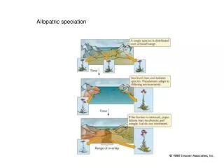
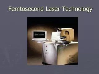
Allopatric Femtosecond Laser Air Bubble Formation in a Closed System. Dan Driscoll, MD Takeshi Ide, MD, PhD; Sonia H Yoo, MD; Richard K Lee, MD, PhD; Terrence P O'Brien, MD Bascom Palmer Eye Institute Miami, FL. Background.

E N D
Allopatric Femtosecond Laser Air Bubble Formation in a Closed System Dan Driscoll, MD Takeshi Ide, MD, PhD; Sonia H Yoo, MD; Richard K Lee, MD, PhD; Terrence P O'Brien, MD Bascom Palmer Eye InstituteMiami, FL Takeshi Ide, MD: research for KANEKA O'Brien, Terrence, MD: consultant for AMO, Alcon, Allergan, B&L, Inspire Pharmaceuticals, Ista Pharmaceuticals Sirion Therapeutics, and Vistakon Pharmaceuticals Sonia Yoo, MD: consultant for Alcon; research for Carl Zeiss Meditec; travel expenses by IntraLase Corpation None of the authors have a propriety interest in this study.
Background • Allopatric air bubble formation happens rarely in femtosecond laser-assisted surgeries • Potential complications of air bubbles: • Impaired suction of laser applanator • Poor intraoperative pachymetry • Poor ablation efficiency • Difficulty with eye tracking and iris registration for excimer laser
Traditionally-held hypothesis for formation • Possibly from air traveling via an intralamellar network to various intraocular locations: • trabecular meshwork • corneal stroma • endothelium • Possibly migration of small bubbles through the posterior stroma and endothelium without being absorbed by the endothelial pump
Materials and Methods • Balanced Salt Solution (BSS) bottles completely filled and capped, verified not to contain air • Optical Coherence Tomography (OCT) was used to determine the thickness of the BSS bottle wall. • Thickness determined to be 500-550 µm
Materials and Methods • Single drop of BSS applied to the bottle surface • Glass applanator then applied without suction ring • 5 bottles sampled for IntraLase flap mode cuts • Flap Settings: 8.5 mm diameter, 180 µm depth, 1.9 µJ bed energy, 2.3 µJ sidecut energy, 11 µm spot separation, 9 µm line separation, 70 degree sidecut angle • Results photographed and measured with Visante OCT
Results • Many bubbles formed in the BSS drop on the surface of the bottle in the areas peripheral to the applanated zone. • During the laser cutting into the bottle wall, many tiny air bubbles gradually appeared inside the BSS bottle. • No cuts crossed the inner wall of the BSS bottles.
In-the-Bottle Air bubble. The air bubbles could be seen clearly under the microscope
OCT image of BSS bottle. IntraLase cut lines in the wall of BSS bottle could be seen clearly under the OCT and no cut crossed the inner wall of the BSS bottles.
Discussion • No observations clinically of air bubbles in the deep stroma or outside the flap in vivo. • Lends discourse to air traveling through the trabecular meshwork, corneal stroma, and endothelium. • Additionally, bubbles initially form centrally and not adjacent to lamellar pocket, which would be expected if TM theory were true. • Bubbles were consistently able to be produced in the closed BSS bottle system with a deeper cut (180 µm) but were not able to be produced with a 120 µm cut.
Proposed mechanism of air bubble formation in closed system • Production allopatric cavitation bubbles in the eye from the vibration of cutting and suction pressure • Similar to cavitation bubbles formed from phacoemulsification • Actual physics behind mechanism still under investigation
Conclusion • Experimental system of BSS bottles does not equal in vivo characteristics of the human eye. • Further research necessary into determining exact mechanism of air bubble formation during femtosecond laser application to a closed system.
References and Acknowledgements Primary investigator for this project was Takeshi Ide, MD, PhD under the direction of Sonia Yoo, MD of the Bascom Palmer Eye Institute 1. Nordan LT, Slade SG, Baker RN, Suarez C, Juhasz T, Kurtz R. Femtosecond laser flap creation for laser in situ keratomileusis: six-month follow-up of initial U.S. clinical series. J Refract Surg. 2003;19:8-14. 2. Seiler T, Koufala K, Richter G. Iatrogenic keratectasia after laser in situ keratomileusis. J Refract Surg. 1998;14:312-317. 3. Genth U, Mrochen M, Walti R, Salaheldine MM, Seiler T. Optical low coherence reflectometry for noncontact measurements of flap thickness during laser in situ keratomileusis. Ophthalmology. 2002;109:973-978. 4. Probst LE, Machat JJ. Mathematics of laser in situ keratomileusis for high myopia. J Cataract Refract Surg. 1998;24:190-195. 5. Seider MI, Ide T, Kymionis GD, Culbertson WW, O'Brien TP, Yoo SH. Epithelial breakthrough during IntraLase flap creation for laser in situ keratomileusis. J Cataract Refract Surg. 2008;34:859-63. 6. Kaiserman I, Maresky HS, Bahar I, RootmanDS.Incidence, possible risk factors, and potential effects of an opaque bubble layer created by a femtosecond laser. J Cataract Refract Surg. 2008;34:417-23. 7. Ide T, Kymionis GD, Goldman DA, Yoo SH, O’Brien TP. Subconjunctival Gas Bubble Formation During LASIK Flap Creation Using Femtosecond Laser. J Refract Surg IN 8. Kuo AN, Kim T. Persistent anterior chamber gas bubbles during IntraLASIK. J Cataract Refract Surg. 2007;33:1134-5. 9. Lifshitz T, Levy J, Klemperer I, LevingerS.Anterior chamber gas bubbles after corneal flap creation with a femtosecond laser. J Cataract Refract Surg. 2005;31:2227-9. 10. Wright JR. Gas bubbles after flap creation with femtosecond laser. J Cataract Refract Surg. 2006;32:1076. 11. Srinivasan S, Rootman DS. Anterior chamber gas bubble formation during femtosecond laser flap creation for LASIK. J Refract Surg. 2007;23:828-30. 12. Cumberbatch E. Self-focusing in Non-linear Optics IMA J Appl Math 1970; 6: 250-262; 13. Mourou GA, Tajima T, Bulanov SV. Optics in the relativistic regime. Rev. Mod. Phys. 2006;78: 309–371. 14. Zacharias J. Role of cavitation in the phacoemulsification process. J Cataract Refract Surg. 2008 May;34:846-52.