Download
1 / 32
320 likes | 331 Views
93 RAYNAUD’S SYNDROME: VASOSPASTIC AND OCCLUSIVE ARTERIAL DISEASE INVOLVING THE DISTAL UPPER EXTREMITY. Vascular Surgery Stanford Hospital and Clinics 02-13-2006. DEFINITION.

E N D
93 RAYNAUD’S SYNDROME: VASOSPASTIC AND OCCLUSIVE ARTERIAL DISEASE INVOLVING THE DISTAL UPPER EXTREMITY Vascular Surgery Stanford Hospital and Clinics 02-13-2006
DEFINITION • Raynaud’s Syndrome – episodic pallor or cyanosis of the fingers due to vasoconstriction of small arteries or arterioles in the fingers occurring in in response to cold or emotional stress • Raynaud’s disease – primary vasospastic disorder without identifiable underlying cause • Raynaud’s phenomenon – vasospasm secondary to an underlying condition or disease
CLINICAL PRESENTATION • Induced by cold exposure • Sudden onset of waxy pallor of digits • Cyanosis follows the pallor • Resolving with hyperemia and rubor of the skin • Female > male (4:1 to 1.6:1)
PREVALENCE • Common – 3.5-4.6% (US) • Higher in cold climates
DIAGNOSIS OF PRIMARY RAYNAUD’S SYNDROM • Vasospastic attacks precipitated by exposure to cold or emotional stimuli • Symmetrical or bilateral involvement of the extremities • Absence of gangrene • Symptom present for a minimum of 2 years • Absence of any other underlying disease
BLOOD FLOW REGULATION OF FINGERS • “Hunting response” – responding to cold temperature, arterial vasoconstriction and dilatation alternates. Frequency about every 30 seconds to 2 minutes
SECONDARY VASOSPASTIC DISORDER • Existing fixed vascular obstruction • Decrease the threshold for cold-induced vasospasm • Conditions causing vessel lumen narrowing - Scleroderma • Increasing viscosity - Myeloma
ANATOMY OF UPPER EXTREMITY AND POTENTIAL ETIOLOGY • Direct compression - Aberrant right subclavian artery, Thoracic outlet syndrome • Embolization – Thoracic outlet syndrome, atherosclerosis • Deep and superficial palmar arches
PHYSICAL EXAMINATION • Investigate causes for secondary Raynaud’s • Exam heart • Upper extremity vascular exams
SEGMENTAL PRESSURE MEASUREMENT • To eval large vessel occlusive diease • Measure systolic pressures at brachial, upper elbow, and wrist • Abnormal – difference > 10 mm Hg • Wrist-brachial ratio - > 0.8
FINGER SYSTOLIC BLOOD PRESSRES • Normal finger-brachial index – 0.8 to 1.27 • Occlusive disease – diff. > 15 mm Hg, or, finger SBP<70 mm Hg • Measure while changing finger temperature
FINGER TIP THERMOGRAPHY • Combined with cold immersion
OTHER TESTS • Cold recovery time – NL <10 mins • Laser Doppler Flux • Duplex ultrasound • Contrast Angiography – gold standard
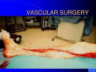
OVERVIEW AND PRESENTATION • Symptomatic UE ischemia is rare – 5% • Most are primary Raynaud’s syndrome – medical management • Acute ischemia – 5 “P”s • Chronic ischemia – equivalent of claudication (dominant hand more) • Tissue loss are rare – rich collaterals • Axillary A. ligation – 10% limb loss • Brachial A. ligation – 3-5% lead to gangrene
ETIOLOGY • Intrinsic arterial disease • Trauma • Iatrogenic • Non-iatrogenic • Embolic
INTRINSIC ARTERIAL DISEASE • Atherosclerosis • Rare to upper extremity • Occasionally seen in axillary, brachial, radial and ulnar A. • FMD • Hypothenar hammer syndrome – distal ulnar A
TRAUMA • Iatrogenic • Brachial A. – most common (0.9-4% after cath) • Axillary A. – 0.8% thrombotic complications • Radial A. – 5-40% (hand ischemia 0.3-0.5%) • Non-iatrogenic • Blunt – intimal disruption, early/late presentation • Traction – intimal disruption (mild), arterial disruption (severe) • Penetrating – direct/blast injury
EMBOLI • Account for 25% total embolic event • External source – cardiac, aortic arch, subclavian A pathology • Intrinsic source – intimal flaps, stenosis, injection • Most common source – cardiac (A-Fib) • Most common location – Brachial A. (60%)
EVALUATION • Acute ischemia – PE • Segmental pressure • Duplex ultrasound • CTA • MRA • Angiogram
TREATMENT • Acute injury – urgent operation • Chronic – depends on clinical presentation
AXILLARY ARTERY • Proximal portion – transverse incision at deltopectoral groove • Distal portion – axillary or upper arm incision • End to end anastomosis • Saphenous vein is the graft of choice • Chronic occlusion – carotid-to-brachial bypass, or axillary-to-brachial bypass
BRACHIAL ARTERY • Embolectomy – incision below the antecubital fossa • Incision right on the projected injury site • Long segment occlusion – Saphenous vein graft • Direct end-to end anastomosis
RADIAL AND ULNAR ARTERIES • Rarely necessary • Acute traumatic injury – urgent repair • Embolectomy – antecubital fossa