Download
1 / 86
880 likes | 905 Views
Dive into the world of sensory receptors and pathways from sensation to perception, exploring various receptor types, adaptation, and pain pathways in a comprehensive overview. Discover how the brain interprets sensory impulses and creates sensations.

E N D
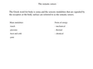
Chapter 12Somatic and Special Senses Somatic (General) Senses • receptors that are widely distributed throughout the body • skin, various organs and joints Special Senses • specialized receptors confined to structures in the head • eyes, ears, nose, mouth
Senses Sensory Receptors • specialized cells or multicellular structures that collect information from the environment • stimulate neurons to send impulses along sensory fibers to the brain Sensation • a feeling that occurs when brain becomes aware of sensory impulse Perception • a person’s view of the stimulus; the way the brain interprets the information
Receptor Types - General Chemoreceptors • respond to changes in chemical concentrations Pain receptors • respond to tissue damage Thermoreceptors • respond to changes in temperature
Mechanoreceptors • respond to mechanical forces proprioceptors –tensions of muscles and tendons baroreceptors – blood pressure stretch receptors – inflation in lungs Photoreceptors • respond to light
Sensory Impulses • Stimulation causes a change in membrane potential • Nerve fiberSensory receptors can be ends of nerve fibers or other cells close to them • generates an action potential and an impulse is sent • Peripheral nerves transmit the impulse to the CNS • CNS analyzes and interprets the impulse
Sensations • Feelings that occur when the brain interprets sensory impulses • All nerve impulses are the same • Sensation depends on which area of the • cerebral cortex receives the impulse • Receptors usually respond to specific • stimuli so the brain creates the correct • sensation
Cerebral cortex interprets the impulse to come from the receptors stimulated • Projection allows a person to pinpoint the region of stimulation Ex: eyes see and the nose smells things
Sensory Adaptation • Occurs when sensory receptors are continuously stimulated • As receptors adapt impulses leave at decreasing rates and may stop entirely • Impulses are only triggered if the strength of the stimulus changes Ex: adjusting to bad smell in a room
Somatic (General) Senses • senses associated with skin, muscles, joints, and viscera • three groups • exteroceptive senses – senses associated with body surface; touch, pressure, temperature, pain • visceroceptive senses – senses associated with changes in viscera; blood pressure stretching blood vessels, ingesting a meal • proprioceptive senses – senses associated with changes in muscles and tendons
Touch and Pressure Senses • common in epithelial tissues • simplest receptors • sense itching Senses mechanical forces that deform or displace tissue 3 types • Sensory nerve fibers – found in epithelial tissue associated with touch and pressure • Meissner’s corpuscles – small oval masses of connective tissue found in hairless part of the skin(lips, palms etc.) sense light touch and texture
Pacinian corpuscles – large masses of connective tissue fibers and cells found in subcutaneous tissues stimulated by heavy pressure
Temperature Senses Heat receptors • sensitive to temperatures above 25oC (77o F) • unresponsive to temperature above 45oC (113oF) Cold receptors • sensitive to temperature between 10oC (50oF) and 20oC (68oF) • Both cold and heat receptors rapidly adapt Pain receptors • respond to temperatures below 10oC • respond to temperatures above 45oC
Pain Receptors • Consist of free nerve endings are located in the skin and internal tissues, except the nervous tissue of the brain • Stimulated when tissue is damaged • Usually specific to the type of pain • Adapt very little, if at all
Visceral Pain • pain receptors are the only receptors in viscera whose stimulation produces sensations • pain receptors respond differently to stimulation • not well localized • may feel as if coming from some other part of the body • known as referred pain • an example would be heartburn
Pain Nerve Pathways Chronic pain fibers • C fibers • thin, unmyelinated • conduct impulses more slowly • associated with dull, aching pain • difficult to pinpoint Acute pain fibers • A-delta fibers • thin, myelinated • conduct impulses rapidly • associated with sharp pain • well localized
An event may trigger impulses on both types of fibers • causing a dull sensation • a sharp pain then a dull aching one • aching pain is usually more intense and may worsen over time • Awareness of pain occurs when the impulse reaches the thalamus • Cerebral cortex judges the intensity and locates the source of pain
Midbrain, pons, and medulla oblongata regulate the flow of impulses from the spinal cord • Neuropeptides (endorphins) inhibit pain sensations • enkephalins and endorphins can suppress chronic and acute pain impulses • serotonin stimulates other neurons to release enkephalins
Stretch Receptors • Proprioceptors that sense length and tensions of muscles - 2 main types Muscle spindles • found in muscles near junctions with tendons • responds to a muscle stretching (stretch reflex) • helps maintain the desired position of a limb
Golgi tendon organs • found in tendons close to muscle attachments • stimulated by increased tension • helps maintain posture and protects muscles from being torn away from attachments
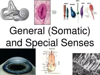
Special Senses • sensory receptors are within large, complex sensory organs in the head • smell in olfactory organs • taste in taste buds • hearing and equilibrium in ears • sight in eyes
Sense of Smell Olfactory Receptors • chemoreceptors • respond to chemicals dissolved in liquids Olfactory Organs • contain olfactory receptors (bipolar neurons) and supporting epithelial cells • cover parts of nasal cavity, superior nasal conchae, and a portion of the nasal septum
Olfactory Nerve Pathways Once olfactory receptors are stimulated, nerve impulses travel through • olfactory nerves olfactory bulbs olfactory tracts limbic system (for emotions) and olfactory cortex in cerebrum(for interpretation) • The only nerve cells in direct contact to the outside environment • the only neurons that are regularly replaced (subject to damage) yet about 1% are lost every year • sense of smell diminishes with age
Olfactory Stimulation • olfactory organs located high in the nasal cavity above the usual pathway of inhaled air • olfactory receptors undergo sensory adaptation rapidly • sense of smell drops by 50% within a second after stimulation Olfactory Code • hypothesis • each odor may stimulate a specific set of receptor subtypes
Sense of Taste Taste Buds • organs of taste • located on papillae of tongue, roof of mouth, linings of cheeks and walls of pharynx Taste Receptors • chemoreceptors • taste cells – modified epithelial cells that function as receptors • taste hairs –microvilli that protrude from taste cells; sensitive parts of taste cells
Taste Sensations Primary Taste Sensations • sweet – most plentiful at tip of tongue • sour – margins of tongue • salty – tip & upper front part of tongue • bitter – back of tongue • umami(savory) – taste of glutamate (MSG) • Food must be dissolved in saliva to be tasted • Taste cells are modified epithelial cells that are replaced continually – taste does not diminish with age
Hearing Ear – organ of hearing Three Sections • External • Middle • Inner
External ear • outer structure called auricle - collects sound waves and directs them inward • external auditory meatus leads inward about 2.5 cm • lined with skin that has modified sweat glands that secrete wax • hairs guard the opening • sound waves pass to the end and alter the pressure on the tympanic membrane (eardrum)
Middle Ear • Includes the tympanic membrane, tympanic cavity, and 3 small bones called auditory ossicles (malleus, incus, stapes) • Tympanic cavity is an air-filled space in the temporal bone that separates the external and internal ears • Tympanic membrane covered by a thin layer of skin on outer surface and mucous membrane on the inside
Auditory ossicles transmit vibrations between the tympanic membrane and inner ear Malleus, incus and stapes • Vibration on the stapes moves fluid within the inner ear which stimulates hearing receptors • Oval window is an opening in the wall of tympanic cavity where the stapes is attached by ligaments
Tympanic reflex reduces pressure from loud sounds that might damage receptors – ossicles become more rigid • Auditory Tube (Eustachian tube) • Connects each middle ear to the throat • Allows air to pass between the tympanic cavity and outside the body
Helps maintain equal pressure on both sides of the tympanic membrane- needed for normal hearing • air pressure changes with altitude causing ears to pop • usually closed by valvelike flaps in the throat • swallowing, yawning, and chewing help open the valves and equalize the pressure
Inner Ear • Complex system of intercommunicating chambers called labyrinth – each ear has 2 regions • osseous labyrinth – bony canal in temporal bone • membraneous labyrinth – a tube within the osseous labyrinth • perylymph is fluid between the 2 chambers
endolymph is fluid in the membraneous labyrinth • cochlea contains the organ of Corti which contains the receptors (hair cells) that vibrations in the fluid stimulates • different frequencies stimulate different receptors • humans can detect sound waves with frequencies from 20 - 20,000 vibrations/second • semicircular canals provide a sense of equilibrium
Auditory Nerve Pathways • Nerve fibers from hearing receptors travel in the cochlear branch of the vestibulocochlear nerves • Impulses travel to the medulla oblongata, midbrain, thalamus, and are interpreted in the temporal lobes of the cerebrum
Sense of Equilibrium Static Equilibrium • Maintains the stability of the head and body when they are motionless • Organs of static equilibrium are the vestibule (bony chamber between the semicircular canals and cochlea)
Sensory receptors are hair cells in a gelatinous material that sense changes in position • Movements tilt the gelatinous mass which stimulates the hair cells to signal nerve fibers • Brain responds by sending impulses to skeletal muscles to contract or relax in order to maintain balance
Macula • responds to changes in head position • bending of hairs results in generation of nerve impulse
Dynamic Equilibrium • Balances the head and body when they are moved or rotated suddenly • Sense organs (hair cells) are located in the ampullae of the semicircular canals • Sudden movements cause the hair cells to bend and send an impulse to the brain • Parts of the cerebellum also interpret information from the semicircular canals and help maintain balance • Visual information is also used to maintain balance