Download
1 / 15
220 likes | 685 Views
The Fetlock and Foot. First Year Anatomy Nicholas Urbanek , BVMS, MRCVS. What is the Fetlock?. Fetlock is the common name for the metacarpophalangeal and metatarsophalangeal joints (MCPJ and MTPJ) of the horses.

E N D
The Fetlock and Foot First Year Anatomy Nicholas Urbanek, BVMS, MRCVS
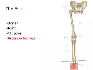
What is the Fetlock? • Fetlock is the common name for the metacarpophalangeal and metatarsophalangealjoints (MCPJ and MTPJ) of the horses. • It is formed by the junction of the third metacarpal (forelimb) or metatarsal (hindlimb) bones (cannon bones) proximally and the proximal phalanx (pastern bone) distally. • Paired proximal sesamoid bones articulate with the palmar or plantar distal surface of the third metacarpal or metatarsal bones and are rigidly fixed to the proximo-palmar /-plantar edge of the proximal phalanx.
Common Views Lateral Lateral DMPLO DP DLPMO
Lateral View • A: Third MC/MT bone • B: Proximal phalanx (P1) • 1: Sagittal ridge • 3: Palmarprocess of proximal phalanx • 4: Condyles of thirdmetacarpalbone • 5: Proximal sesamoidbones
Dorso-palmer/-plantar (DP) • A: Third MC/MT bone • B: Proximal phalanx • C: Medial proximal sesamoidbone • D: Lateral proximal sesamoidbone • E: Metacarpo(tarso)-phalangeal joint • J: Depression for medialcollateral ligament attachment • 1: Sagittal ridge
Obliques – A Review • The view is named for the projection of the beam. • Typically taken at 45 degree angles off the sagittal axis of the limb. • Markers must be in place, otherwise unable to distinguish medial from lateral side. • Dorsomedial-palmarolateral (plantar)oblique (DMPLO) • Dorsolateral-palmaromedial (plantar)oblique (DLPMO)
Dorsal Lateral Medial Palmar/Pl DLPMO A: Third MC/MT bone B: Proximal phalanx C: Medial proximal sesamoid bone D: Lateral proximal sesamoid bone E: Metacarpo(tarso)-phalangeal joint 1: Sagittal ridge 3: Palmar process of proximal phalanx/Lateral palmar tubercle (eminence) 6: Medial condyle of third MC/MT bone DLPMO
Dorsal Lateral Medial Palmar/Pl A: Third MC/MT bone B: Proximal phalanx C: Medial proximal sesamoid bone D: Lateral proximal sesamoid bone E: Metacarpo(tarso)-phalangeal joint 1: Sagittal ridge 2: Lateral condyle of third metacarpal/tarsal bone 3: Palmar process of proximal phalanx DMPLO DMPLO
Fetlock Bulging Forelimb vs Hindlimb • The MT isconvexatits distal aspect DP view - Hindlimb DP view - Forelimb The proximal sesamoidbones are higher The proximal sesamoidbones are more triangular
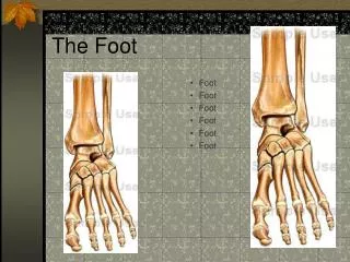
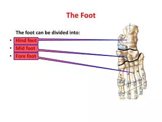
Foot or Distal Limb • Composed of four bones • Proximal, middle, distal (first, second, and third) phalanx • Navicular bone • Multiple views obtained • Lateral • Dorsopalmar(plantar) view • Dorsoproximal-palmar(plantar)odistal oblique • “Upright Pedal” or “High coronary” • Palmar(plantar)oproximal-palmar(plantar)odistal • “Skyline Novicular” • Other oblique views • The foot should have no shoe, be trimmed, and sulci should be packed with Play-Doh • Marker always to the lateral side…can not tell laterality otherwise
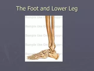
Lateral view A: Middle phalanx B: Third phalanx C: Navicular bone 1: Proximal interphalangeal joint 2: Distal interphalangeal joint 3: Extensor process 4: Dorsal surface 5: Palmar process
A: Middle phalanx B: Distal phalanx C: Navicular bone 6: Proximal interphalangeal joint 7: Distal interphalangeal joint Dorsopalmar(plantar) view R 6 A 7 C B
2 5 5 B 6 7 Dorsoproximal-palmar(pl)odistal oblique view A A: Middle phalanx B: Third phalanx C: Navicular bone 1: Proximal interphalangeal joint 2: Distal interphalangeal joint 3: Extensor process 4: Dorsal surface 5: Palmar process 6: Vascular channel 7: Solar margin R “Upright pedal” “High coronary” Dorsoproximal-palmar(plantar)odistal oblique view
Palmaro(plantar)oproximal-palmar(plantar)odistal oblique view “Skyline Novicular” C: Navicular bone 3: Articular surface 8: Palmar aspect of middle phalanx 9: Nutrient foramen 10: Sagittal ridge 11: Articulation between navicular bone and middle phalanx