Download
1 / 37
410 likes | 765 Views
Animal Locomotory Systems. Biology 2: Form and Function. Types of skeleton. Hydraulic or hydrostatic skeletons use a fixed volume, non-compressible fluid contained within a sack, against which muscular contractions are applied

E N D
Animal Locomotory Systems Biology 2: Form and Function
Types of skeleton • Hydraulic or hydrostatic skeletons use a fixed volume, non-compressible fluid contained within a sack, against which muscular contractions are applied • Exoskeleton surround the body, and are rigid, and often impermeable. As it is non-living, growth of an exoskeleton is problematic • Endoskeleton are rigid internal skeleton made of living connective tissue capable of growth and self-repair
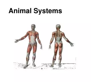
The human skeletal system • Axial skeleton • Skull • Ribs • Spine • Appendicular skeleton • Pelvis • Limbs • Hands • Feet
The structure of bone • Bone consists of brittle but rigid Calcium Phosphate, interweaved with flexible but weak collagen • New bone is made in osteoblasts, intitially as cartilage (non-calcified bone) • New bone cells, osteocytes, are encase within lacunae, and may eventually be reconstituted by osteoclasts • Bone is laid down in layers (lamellae) located around Haversian canals • Ends and interior bone contains a more open lattice (spongy bone) which contains bone marrow
Types of joint • Fixed joint (immovable). e.g, sutures in skull • Slightly movable joints. e.g., spine • Movable (synovial joints) - full range of motion • Ball and socket (pelvis-femur, humerous-scapula) • Hinge (elbow) • Rotating (axis-atlas)
Muscles move bones • Muscle may act together (synergistic) or against each other (antagonistic) • Muscles contract by electrical stimulation from nervous system • Electrical stimulation can be replicated artificially to demonstrate a graded response • Single contraction-stimulation = twitch • Multiple contraction stimulations with slight recovery = summation • Multiple contraction stimulations with no recovery = tetanus • Isotonic contractions are those that result in muscle shortening • If muscle does not shorten because load is too great, then contraction is isometric
Muscle composition • Muscles contain muscle fibres • A group of muscle fibers served by the same neuron is termed a motor unit • Muscle fibres (= cell) contains a bundle of myofibrils • Myofibrils are striated by dark and light bands • Dark bands consist of thick myofibrils • Light bands consist of thin myofibrils • At rest, light bands barely overlap with dark bands
Fine structure of the myofibril • At rest, thin myofilaments and thick myofilaments barely overlap • Light, or I-bands, are separated in their middle by z-lines • Thick, or A-bands, have a lighter center (referred to as an H-band) • Whole unit, from I-band to I-band (including one A-band, is termed a sarcomere, and is the functional contracting unit • On contraction, thin myofilaments slide along, and into the thick myofilaments, shortening the H-and z-lines; termed the sliding filament mechanism of contraction
Contraction at the molecular level • Thick myofilaments consist of myosin fibres • Each myosin fibre has a head that can attach to a thin fibre as a cross-bridge • Thin myofilaments consist of actin fibres that can bind to the myosin heads of a thick myofilament • In the act of muscle contraction, Myosin converts ATP to ADP. This reconfigures the myosin head to bind to a actin fibre. As the myosin fibre contracts, the thin myofilament is dragged with it • At the end of this power stroke, the myosin head binds with further ATP - this releases the cross bridge, allowing the myosin to rebind at a site further up the thin myofilament
The control of contraction • Motor neurons release a neurotransmitter, Acetylcholine (ACh), which prompts the muscle fibre membrane to poduce an electrochemical impulse • Impulses travel along cellular invaginations known as transverse, or t-tubules • t-tubules carry impulse to the sarcoplasmic reticulum, as repository of Ca2+ • Ca2+ binds to troponin, a protein that in combination with tropomysosin, isolates the actin molecules of the thin myofilaments. During contraction, the binding of Ca2+ exposes the thin myofilament to crossbridge formation • Muscle relaxation is prompted by the cessation of nervous stimulation, which inhibits the supply of Ca2+ ions to troponin. The troponin-tropomyosin complex thus returns to its protective role, preventing crossbridge formation
Types of muscle fibres • Slow-twitch (Type I, SlowOxidative, or red) fibres are deep red in color due to high levels of myoglobin, and have a high capacity for aerobic respiration and resist fatigue • Fast-twitch (Type II, Glycolitic, or white) fibres contain less myoglobin, are adapted to anaerobic respiration, and are capable of rapid, non-sustainable generation of power (can be strengthened through exercise) • Intermediate fast-twitch (FastOxidative) fibres are also resistant to fatigue. Performance may be improved by endurance exercises