Download
1 / 25
390 likes | 1.47k Views
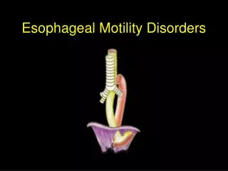
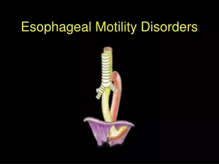
The New Era of Esophageal Motility Disorders. Joint Hospital Surgical Ground Round April 2014 Hwang Wan Wui Winston Queen Elizabeth Hospital. Presentation Outline. Chicago Classification Scheme High Resolution Esophageal Pressure Topography Achalasia Presentation Investigation Treatment.

E N D
The New Era of Esophageal Motility Disorders Joint Hospital Surgical Ground Round April 2014 Hwang Wan Wui Winston Queen Elizabeth Hospital
Presentation Outline • Chicago Classification Scheme • High Resolution Esophageal Pressure Topography • Achalasia • Presentation • Investigation • Treatment
Chicago Classification Scheme • High Resolution Esophageal Pressure Topography • Achalasia • Presentation • Investigation • Treatment
The Chicago Classification • Investigators in the Northwestern University in Chicago developed a new classification scheme to facilitate the diagnosis of esophageal motility disorders by interpretation of high resolution esophageal pressure topography (EPT) • Esophageal pressure topography is a combination of high resolution manometry (HRM) and pressure topography
The Chicago Classification IRP Integrated Relaxation Pressure Each metric developed to characterize a specific feature of deglutitive esophageal function
Esophageal Pressure Topography V.S. Conventional Manometry EPT allows more sophisticated interpretation of esophageal motility
The Chicago Classification Achalasia Type I: Classic Type II: Pan-esophageal pressurization Type III: Spastic Achalasia Type I: Classic Type II: Pan-esophageal pressurization Type III: Spastic IRP >= upper limit of normal AND absent peristalsis IRP >= upper limit of normal AND absent peristalsis YES NO IRP >= upper limit of normal AND some instances of intact or weak peristalsis IRP >= upper limit of normal AND some instances of intact or weak peristalsis YES EGJ Outflow Obstruction Achalasia variant Mechanical obstruction EGJ Outflow Obstruction Achalasia variant Mechanical obstruction NO IRP is normal BUT abnormalities in other metrics IRP is normal BUT abnormalities in other metrics Other Esophageal Motility Disorders Distal esophageal spasm Hypercontractile esophagus Absent peristalsis Nutcracker esophagus YES Other Esophageal Motility Disorders Distal esophageal spasm Hypercontractile esophagus Absent peristalsis Nutcracker esophagus
Chicago Classification Scheme • High Resolution Esophageal Pressure Topography • Achalasia • Presentation • Investigation • Treatment
Achalasia - Presentation • Dysphagia • both liquid and solid • Regurgitation • Chest pain • Cough • Aspiration pneumonia • Weight loss
Achalasia - Investigations • Barium Esophagogram • Classical “bird’s beak” appearance • Dilated esophageal body • High Resolution Manometry • Esophagogastroduodenoscopy • Rule out pseudoachalasia • Most common cause is malignancy infiltrating the EGJ
Achalasia – Type I (Classic Achalasia) • Mean IRP >= upper limit of normal (IRP =42mmHg) • 100% failed peristalsis
Achalasia – Type II • Mean IRP >= upper limit of normal • No normal peristalsis • Panesophageal pressurization with >20% of swallows, which may exceed LES pressure, causing the esophagus to empty
Achalasia – Type III (Spastic Achalasia) • Mean IRP >= upper limit of normal • No normal peristalsis • Fragments of premature (spastic) distal contractions with 20% of swallows • Although this is also associated with rapidly propagated pressurization, the pressurization is attributable to an abnormal lumen obliterating contraction
Achalasia TreatmentPharmacological • Calcium channel blockers and nitrates • short lived response • side effects: headache, dizziness and pedal edema • Botulin toxin injection • prevents the release of acetylcholine at terminal nerve endings • results last 6-9 months. [1] • Pharmacological therapies are less effective than endoscopic or surgical therapies [1] Pasricha PJ, Ravich WJ, Hendrix TR, Sostre S, Jones B, Kal- loo AN. Intrasphincteric botulinum toxin for the treatment of achalasia. N Engl J Med 1995; 332: 774-778
Achalasia TreatmentPneumatic Dilation • Aims at disrupting the LES by forceful dilation using air filled balloons • Many use a graded dilation protocol starting with 3.0 cm, then stepping up to 3.5cm and 4.0cm
Achalasia TreatmentPneumatic Dilation • Promising short term results • Long term follow-up showed recurrence [1] Eckardt VF, Gockel I, Bernhard G. Pneumatic dilation for achalasia: late results of a prospective follow up investigation. Gut 2004; 53: 629-633 [2] Katsinelos P, Kountouras J, Paroutoglou G, Beltsis A, Zavos C, Papaziogas B, Mimidis K (2005) Long-term results of pneu- matic dilation for achalasia: a 15 years’ experience. World J Gastroenterol 11:5701–5705
Achalasia TreatmentLaparoscopic Heller’s Myotomy (LHM) • Myotomy from 1.5-3cm distal to the EGJ dividing the longitudinal and oblique muscle to 6-8cm proximal to the EGJ dividing longitudinal and circular muscle of esophagus • Partial fundoplication is routinely performed as incidence of reflux after Heller’s myotomy is >50%
Achalasia TreatmentLaparoscopic Heller’s Myotomy (LHM) • LHM considered superior to pneumatic dilation and the first choice of treatment for achalasia • Prospective trials have shown promising long term results of LHM • A prospective trial in Italy followed up 6 years after laparoscopic Heller-Dor operation [1] • Primary outcome was therapeutic success in terms of symptoms improvement • At 6 years, 81.7% of patients still have significant improvement in their symptoms [1] Costantini M, Zaninotto G, Guirroli E, et al. The laparoscopic Heller-Dor operation remains an effective treatment for esophageal achalasia at a minimum 6-year follow-up. Surg Endosc 2005;19:345-51
Achalasia TreatmentLaparoscopic Heller’s Myotomy (LHM) • Multicenter RCT published by European Achalasia Trial group in 2011 [1] • Primary outcome was therapeutic success, measured by Eckardt score • After 2 years of follow up, the study concluded LHM was notsuperior to pneumatic dilation • Limitations: • 2 year cohort study with no evidence on intermediate and long-term remission rates • All patients in the PD group received 2 to 3 sessions of redilation [1] Boeckxstaens GE, Annese V, des Varannes SB, Chaussade S, Costantini M, Cuttitta A, Elizalde JI, Fumagalli U, Gaudric M, Rohof WO, Smout AJ, Tack J, Zwinderman AH, Zaninotto G, Busch OR. Pneumatic dilation versus laparoscopic Heller’ s myotomy for idiopathic achalasia. N Engl J Med 2011; 364: 1807-1816
Achalasia Treatment in Different Subtypes [1] Pandolfino JE, Kwiatek MA, Nealis T, Bulsiewicz W, Post J, Kahrilas PJ. Achalasia: a new clinically relevant classification by high-resolution manometry. Gastroenterology 2008;135:1526-1533. [2] Salvador R, Costantini M, Zaninotto G, et al. The preoperative manometric pattern predicts the outcome of surgical treatment for esophageal achalasia. J Gastrointest Surg 2010;14:1635–1645 [3] Rohof WO, Salvador R, Annese V, et al. Outcomes of treatment for achalasia depend on manometric subtype. Gastroenterology 2013; 144:718–725
Peroral Endoscopic Myotomy (POEM) • Dissection of inner circular muscle layer of the esophagus • Dissection begins around 7cm proximal to EGJ and down to 2cm distal to EGJ • Good short-term results • Long-term results not available yet
Conclusion • Pharmacological therapies are not recommended unless patient is not fit for endoscopic or surgical therapies • Pneumatic dilation is the most effective nonsurgical treatment with promising short term results but high recurrence rate in the long term • Laparoscopic Heller’s myotomy should be advocated for patients fit for surgery • The Chicago Classification Scheme is providing a better classification for esophageal motility disorders. It has great impact on how we approach esophageal motility disorders, predict treatment outcomes and choose treatment options
The New Era of Esophageal Motility Disorders Winston Hwang Queen Elizabeth Hospital
References • Goldblum JR, Rice TW, Richter JE. Histopathologic features in esophagomyotomy specimens from patients with achala- sia. Gastroenterology 1996; 111: 648-654 • Richter JE. Achalasia – An Update. J Neurogastroenterol Motil. Jul 2010; 16(3): 232–242 • Boeckxstaens GE, etal. Achalasia. Lancet 2014; 383: 83-93 • Stefanidis D, et al. SAGES guidelines of the surgical treatment of esophageal achalasia. Surg Endosc 2012; 26: 296-311