Download
1 / 57

E N D
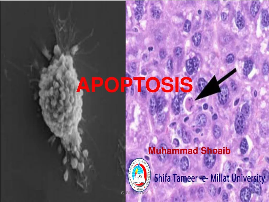
APOPTOSIS Muhammad Shoaib G. PATHOLOGY
Discussion headings • Introduction • Etiopathogenesis • Morphological, Biochemical changes • Mechanism – Intrinsic & Extrinsic pathway • Disorders of apoptosis • Conclusion G. PATHOLOGY
Introduction G. PATHOLOGY
Apoptosis - Definition • A pathway of cell death induced by a tightly regulated suicidal program, in which the cells destined to die activate enzymes that degrade cells own nuclear DNA and nuclear, cytoplasmic proteins. G. PATHOLOGY
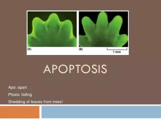
The word "apoptosis" (ἁπόπτωσισ) is used in Greek to describe the "dropping off" or "falling off" of petals from flowers, or leaves from trees. the second half of the word being pronounced like "ptosis" (with the "p" silent), which comes from the same root "to fall", and is already used to describe the drooping of the upper eyelid Kerr Wyllie and Currie paper, British Journal of Cancer, 1972 Aug;26(4):239-57 G. PATHOLOGY
Historical aspects • German scientist Carl Vogt - Principle of apoptosis (1842). G. PATHOLOGY
Walther Flemming – Process of programmed cell death (1845). G. PATHOLOGY
John Foxton Ross Kerr – Distinguish apoptosis from traumatic cell death (1962). G. PATHOLOGY
Cell death mechanisms Death by suicide Death by injury G. PATHOLOGY
Significance of apoptosis • During development many cells are produced in excess which eventually undergo programmed cell death and thereby contribute to sculpturing many organs and tissues [Meier, 2000] • In human body about one lakh cells are produced every second by mitosis and a similar number die by apoptosis (Vaux and Korsmayer ,1999, cell) • Between 50 and 70 billion cells die each day due to apoptosis in the average human adult. For an average child between the ages of 8 and 14, approximately 20 billion to 30 billion cells die a day. (Karam, Jose A. (2009). Apoptosis in Carcinogenesis and Chemotherapy. Netherlands: Springer. ISBN978-1-4020-9597-9) G. PATHOLOGY
Etiopathogenesis G. PATHOLOGY
Why should a cell commit suicide? • 1. Programmed cell death is as needed for proper normal development as mitosis is. Examples: • The resorption of the tadpole tail in frog . • The formation of the fingers and toes of the fetus requires the removal, by apoptosis. • The sloughing off of the endometrium at the start of menstruation. • The formation of the proper connections (synapses) between neurons in the brain. G. PATHOLOGY
2. Programmed cell death is needed to destroy cells that represent a threat to the integrity of the organism. • Examples: • Cells infected with viruses • Cells of the immune system • Cells with DNA damage • Cancer cells (Uncontrolled proliferated cells) G. PATHOLOGY
Apoptosis in physiologic situations • Programmed destruction during embryogenesis • Involution of hormone dependent tissues • Cell loss in proliferating cell populations • Elimination of harmful self- reactive lymphocytes • Death of host cells G. PATHOLOGY
Apoptosis in bud formation during which many interdigital cells die. They are stained black by a TUNEL method Incomplete differentiation in two toes due to lack of apoptosis G. PATHOLOGY
Apoptosis: in embryogenesis Morphogenesis (eliminates excess cells): Selection (eliminates non-functional cells): G. PATHOLOGY
Apoptosis: in embryogenesis Immunity (eliminates dangerous cells): Self antigen recognizing cell Organ size (eliminates excess cells): G. PATHOLOGY
Apoptosis: importantance in adults Tissue remodeling (eliminates cells no longer needed): Apoptosis Involution (non-pregnant, non-lactating) Late pregnancy, lactation Virgin mammary gland - Testosterone Apoptosis Prostate gland G. PATHOLOGY
Apoptosis: importantance in adults Tissue remodeling (eliminates cells no longer needed): Apoptosis + antigen (e.g. infection) - antigen (e.g. recovery) Resting lymphocytes G. PATHOLOGY
Apoptosis in pathological conditions - DNA damage - Accumulation of misfolded proteins - Cell death in certain infections - Pathological atrophy in parenchymal organs G. PATHOLOGY
Cells of the immune system • CTLs induce apoptosis in each other and even in themselves. • Defects in the apoptotic machinery is associated with autoimmune diseases such as lupus erythematosus and rheumatoid arthritis. G. PATHOLOGY
Cells infected with viruses • One of the methods by which cytotoxic T lymphocytes (CTLs) kill virus-infected cells is by inducing apoptosis G. PATHOLOGY
Cells with DNA damage • Damage to its genome can cause a cell • to disrupt proper embryonic development leading to birth defects • to become cancerous. • Cells respond to DNA damage by increasing their production of p53. p53 is a potent inducer of apoptosis. G. PATHOLOGY
Cancer cells • Radiation and chemicals used in cancer therapy induce apoptosis in some types of cancer cells. Fig. 1: SC-1 induced apoptosis in stomach carcinoma cells Left: Before induction Middle: 24h after induction Right: 48h after induction G. PATHOLOGY
Morphological & Biochemical changes G. PATHOLOGY
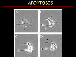
Classic changes • Cell shrinkage • Nuclear fragmentation • Chromatin condensation • Chromosomal DNA fragmentation • Formation of cytoplasmic blebs& apoptotic bodies • Phagocytosis G. PATHOLOGY
Bio chemical changes G. PATHOLOGY
Mechanisms of apoptosis G. PATHOLOGY
Caspases • Caspase are Cysteine- Aspartic acid specific proteases that mediates the events that are associated with programmed cell death. • Their catalytical activity depends on a critical cysteine-residue within a highly conserved active-site pentapeptide QACRG, • Caspases specifically cleave their substrates after Asp residues. • Caspase- 8, Caspase- 9 • acts as an initiator of the caspase activation cascade. • Caspase-3 • key effector in the apoptosis pathway, amplifying the signal from initiator caspases and signifying full commitment to cellular disassembly. G. PATHOLOGY
Initiation • Absence of stimuli - hormones, growth factors • Activation of receptors – TNF family • Heat ,radiation, chemicals • Genetically programmed events G. PATHOLOGY
STAGES OF CLASSIC APOPTOSIS Healthy cell DEATH SIGNAL / STIMULI (extrinsic or intrinsic) Commitment to die (reversible) EXECUTION (irreversible) Dead cell (condensed, crosslinked) ENGULFMENT (macrophages, neighboring cells) DEGRADATION G. PATHOLOGY
Receptor pathway (physiological): FAS ligand TNF Death receptors: (FAS, TNF-R, etc) Death domains Adaptor proteins Pro-caspase 8 (inactive) Caspase 8 (active) Pro-execution caspase (inactive) Execution caspase (active) MITOCHONDRIA Death G. PATHOLOGY
Apoptosis triggered by external signals: the extrinsic or death receptor pathway G. PATHOLOGY
Regulation of apoptosis • Regulatory proteins – BCL -2, equivalent to CED -9 • Apoptosis depends on binding of BCL -2 with pro apoptotic and anti apoptotic proteins. • Situated in the outer mitochondrial membrane. • Apaf -1 equivalent to CED -4. • Tp 53, caspases, BAX, viruses such as adeno, papilloma , hepatitis B. G. PATHOLOGY
Intrinsic pathway (damage): Mitochondria Cytochrome c release BAX BAK BOK BCL-Xs BAD BID B IK BIM NIP3 BNIP3 BCL-2 BCL-XL BCL-W MCL1 BFL1 DIVA NR-13 Several viral proteins Pro-caspase 9 cleavage Pro-execution caspase (3) cleavage Caspase (3) cleavage of cellular proteins, nuclease activation, etc. Death G. PATHOLOGY
Intracellular signals Oxidative damage from free radicals, Radiation, Virus infection, Nutrient deprivation, Pro-apoptotic Factors Damage to the mitochondrial membrane increasing permeability Entry of Cytochrome C into the cytoplasm Cytochrome C binds to Apaf-1 forming an apoptosome Apoptosome activates procaspase-9 to caspase-9 Caspase-9 cleaves and activates caspase-3 and caspase-7. This executioner caspases activate a cascade of proteolytic activity that leads to: Chromatin condensation, DNA fragmentation, Protein cleavage, Membrane permeability G. PATHOLOGY
Physiological Intrinsic receptor pathway damage pathway MITOCHONDRIAL SIGNALS Caspase cleavage cascade Orderly cleavage of proteins and DNA CROSSLINKING OF CELL CORPSES; ENGULFMENT (no inflammation) G. PATHOLOGY
Steps G. PATHOLOGY
Disorders of apoptosis G. PATHOLOGY
Apoptosis: Role in Disease TOO MUCH: Tissue atrophy Neurodegeneration Thin skin etc TOO LITTLE: Hyperplasia Cancer Athersclerosis etc G. PATHOLOGY
Apoptosis: Role in Disease Neurodegeneration • Neurons are post-mitotic (cannot replace themselves; neuronal stem cell • replacement is inefficient) • Neuronal death caused by loss of proper connections, loss of proper growth • factors (e.g. NGF), and/or damage (especially oxidative damage). • Neuronal dysfunction or damage results in loss of synapses or loss of cell • bodies (synaptosis, can be reversible; apopsosis, irreversible) • PARKINSON'S DISEASE • ALZHEIMER'S DISEASE • HUNTINGTON'S DISEASE etc. G. PATHOLOGY
Apoptosis: Role in Disease Cancer • Apoptosis eliminates damaged cells (damage => mutations => cancer • Tumor suppressor p53 controls senescence and apoptosis responses to damage. • Most cancer cells are defective in apoptotic response(damaged, mutant cells survive) • High levels of anti-apoptotic proteins • or • Low levels of pro-apoptotic proteins ===> CANCER G. PATHOLOGY
Apoptosis: Role in Disease Cancer Virus associated cancer • Several human papilloma viruses(HPV) have been implicated in causing cervical cancer. One of them produces a protein (E6) that binds and inactivates the apoptosis promoter p53. • Epstein-Barr Virus (EBV), the cause of mononucleosis and associated with some lymphomas • produces a protein similar to Bcl-2 • produces another protein that causes the cell to increase its own production of Bcl-2. Both these actions make the cell more resistant to apoptosis (thus enabling a cancer cell to continue to proliferate). G. PATHOLOGY
Apoptosis: Role in Disease Cancer Some B-cell leukemia and lymphomas express high levels of Bcl-2, thus blocking apoptotic signals they may receive. The high levels result from a translocation of the BCL-2 gene into an enhancer region for antibody production. Melanoma (the most dangerous type of skin cancer) cells avoid apoptosis by inhibiting the expression of the gene encoding Apaf-1. G. PATHOLOGY