Download
1 / 20
260 likes | 721 Views
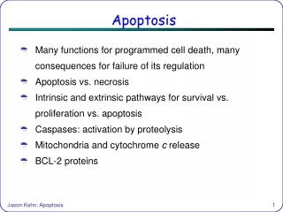
APOPTOSIS. Dr Sarah Meachem. Intentions of this talk. Define apoptosis terminology, methods to detect Define necrosis terminology Understand the differences between apoptosis & necrosis. Importance of Apoptosis. Programmed cell death Regulate cell numbers eg spermatogenesis

E N D
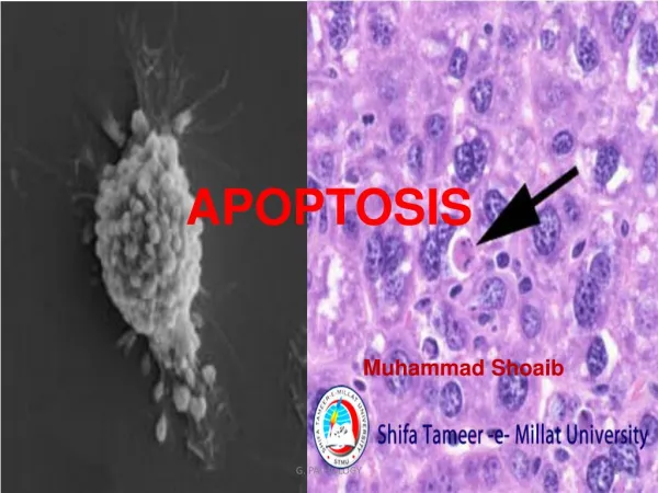
APOPTOSIS Dr Sarah Meachem
Intentions of this talk • Define apoptosis terminology, methods to detect • Define necrosis terminology Understand the differences between apoptosis & necrosis
Importance of Apoptosis Programmed cell death • Regulate cell numbers eg spermatogenesis • Facilitate morphogenesis eg interdigital tissue • Remove potentially harmful cells eg self-reactive T-cells • Eliminate no longer needed cells eg tadpole tail
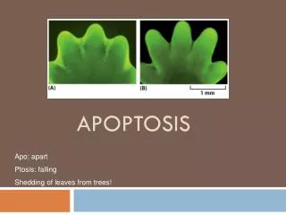
A classic example of apoptosis • Interdigitation see
When programmed cell death goes wrong! So PCD/apoptosis is a normal part of healthy development
Why, what & how of Apoptosis! • Why do they do it? Developmental plasticity • What tells them to do it? Specific physiological signals • How do they do it? Activation of cell death genes
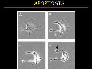
Hallmarks of Apoptosis • Membrane blebbing (photo d) • Chromatin condensation • DNA fragmentation • Phagocytic removal of dead cells From Nicholas et al Nature 2001
Necrosis vs apoptosis…morphological features • Membrane blebbing, but no loss of integrity • Aggregation of chromatin at the nuclear membrane • Cellular condensation (shrinkage) • Formation of membrane bound vesicles (apoptotic bodies) • No disintegration of organelles, organelles remain intact, mitochondria become leaky • Loss of membrane integrity • Begins with swelling of cytoplasm & mitochondria • Swelling of cell and lysis • No vesicle formation, complete lysis • Disintegration of organelles
Necrosis vs apoptosis…biochemical features • Loss of regulation of ion homeostasis • No energy requirement (passive process), also occurs at 4C • Random digestion of DNA • Postlytic DNA fragmentation (late event of death) • Tightly regulated process involving activation & enzymatic steps • Energy (ATP)-dependent active process, does not occur at 4C • Non random mono/oligonucleosomal length fragmentation of DNA • Prelytic DNA fragmentation • Activation of the caspase cascade
Necrosis vs apoptosis…physiological significance • Death of cell groups • Evoked by non-physiological disturbances • Phagocytosis by macrophages • Significant inflammatory response • Death of single (individual) cells • Induced by physiological stimuli • Phagocytosis by adjacent cells or macrophages • No inflammatory response
FasL Extrinsic Pathway (Death receptor) Fas D D D D D Intrinsic Pathway (Mitochondrial) FADD Endoplasmic Reticulum Pathway Procaspase 8 Caspase 8 Apaf-1 Cytochrome C Procaspase 9 Caspase 12 Caspase 3, 6 & 7 Apaf-1 Caspase 9 APOPTOSIS Caspase cascade-3 apoptotic pathways
How to identify apoptosis • Morphology (light/electron microscopy) • DNA laddering (isolated cells) • In situ detection of DNA fragmentation (fixed tissues) • Annexin V- protein • Activated caspase activity (proteins)
How to identify apoptosis…cont TUNEL Caspase 9 Co-localisation Normal men Gonadotrophin deplete men (2 weeks) Caspase 8
Causes of cell injury…. External forces • Lack of oxygen • Physical agents (trauma, burn, freeze, radiation, electricity) • Chemical agents (drugs, poisons, toxins, heavy metals) • Infectious agents (bacteria, viruses, parasites) Internal factors • Immunologic • Genetic • Metabolic
Types of necrosis • Coagulative –infarction- heart (common, denaturations of proteins) • Liquifactive- brain, abscess • Caseous-tuberculosis • Gangrene-with infection-limbs
Cerebral infarct liquefactive necrosis; fluid-filled space is formed in necrotic area.
Extensive Caseous necrosisTuberculosis V. Shashidar, Fiji School of Medicine
Gangrene - Diabetic foot V. Shashidar, Fiji School of Medicine