Download
1 / 34
350 likes | 387 Views
Learn about the fundamentals of joints, including their classification into structural and functional categories, and the different types of joints such as fibrous, cartilaginous, and synovial. Understand how joints provide mobility and stability to the skeleton.

E N D
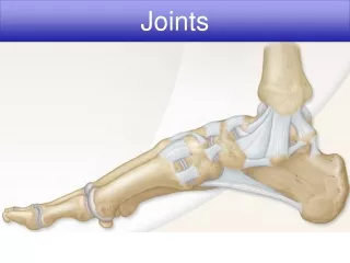
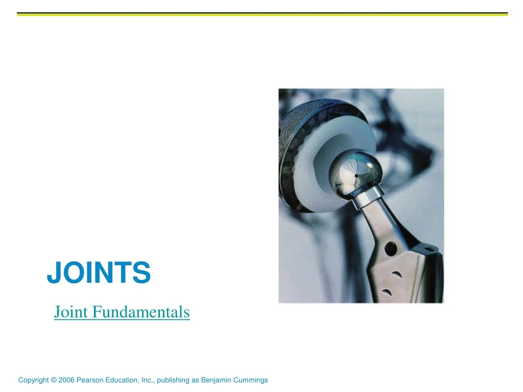
Joints Joint Fundamentals
Joints (Articulations) • Weakest parts of the skeleton • Articulation – site where two or more bones meet • Functions of joints • Give the skeleton mobility • Hold the skeleton together
Classification of Joints: Structural • Structural classification focuses on the material binding bones together and whether or not a joint cavity is present • The three structural classifications are: • Fibrous • Cartilaginous • Synovial
Classification of Joints: Functional • Functional classification is based on the amount of movement allowed by the joint • The three functional classes of joints are: • Synarthroses – immovable • Amphiarthroses – slightly movable • Diarthroses – freely movable
Fibrous Structural Joints • The bones are joined by dense, connective fibrous tissues • There is no joint cavity • Most are immovable • There are three types – sutures, syndesmoses, and gomphoses
Fibrous Structural Joints: Sutures • Occur between the bones of the skull • Comprised of interlocking junctions completely filled with connective tissue fibers • Bind bones tightly together, but allow for growth during youth • In middle age, skull bones fuse and are called synostoses
Fibrous Structural Joints: Sutures Figure 8.1a
Fibrous Structural Joints: Syndesmoses • Bones are connected by a fibrous tissue ligament • Movement varies from immovable to slightly variable • Examples include the connection between the tibia and fibula, and the radius and ulna
Fibrous Structural Joints: Syndesmoses Figure 8.1b
Review • Name two functions of joints. • Name three structural classification of joints. • Name three functional classification of joints. • Name three types of fibrous joints. • Where is the most prominent place to find suture joints. • Name one example of Syndesmoses joint. Joint Fundamentals
Fibrous Structural Joints: Gomphoses • The peg-in-socket fibrous joint between a tooth and its alveolar socket • The fibrous connection is the periodontal ligament
Cartilaginous Joints • Articulating bones are united by hyaline cartilage or disks of fibrocartilage • Lack a joint cavity • Two types – synchondroses and symphyses
Cartilaginous Joints: Synchondroses • A bar or plate of hyaline cartilage unites the bones • Examples include: • Epiphyseal plates of children • Joint between the costal cartilage of the first rib and the sternum
Cartilaginous Joints: Synchondroses Figure 8.2a, b
Cartilaginous Joints: Symphyses • Hyaline cartilage covers the articulating surface of the bone and is fused to an intervening pad of fibrocartilage • Amphiarthrotic joints designed for strength and flexibility • Examples include intervertebral joints and the pubic symphysis of the pelvis
Cartilaginous Joints: Symphyses Figure 8.2c
Synovial Joints • Those joints in which the articulating bones are separated by a fluid-containing joint cavity • Ends of the bones are covered in hyaline cartilage and the ends are encapsulated by synovial fluid • All are freely movable • Examples – all limb joints, and most joints of the body
Synovial Joints: General Structure • Synovial joints all have the following • Articular cartilage • Joint (synovial) cavity • Articular capsule • Synovial fluid • Reinforcing ligaments
Synovial Joints: General Structure Figure 8.3a, b
Ball-and-Socket Joints Types of Synovial Joints • A spherical or hemispherical head of one bone articulates with a cuplike socket of another • Multiaxial joints permit the most freely moving synovial joints • Examples: shoulder and hip joints
Ball-and-Socket Joints Figure 8.7f
Condyloid Joints • Oval articular surface of one bone fits into a complementary depression in another • Both articular surfaces are oval • Biaxial joints permit all angular motions • Examples: radiocarpal (wrist) joints, and metacarpophalangeal (knuckle) joints
Condyloid Joints Figure 8.7d
Planar Joints • One flat bone surface glides or slips over another similar surface • Examples – intercarpal and intertarsal joints, and between the flat articular processes of the vertebrae
Planar Joints Figure 8.5a
Hinge Joints • Hinge joints • Cylindrical projections of one bone fits into a trough-shaped surface on another • Motion is along a single plane • Uniaxial joints permit flexion and extension only • Examples: elbow and interphalangeal joints
Hinge Joints Figure 8.7b
Pivot Joints • Rounded end of one bone protrudes into a “sleeve,” or ring, composed of bone (and possibly ligaments) of another • Only uniaxial movement allowed • Examples: joint between the axis and the dens, and the proximal radioulnar joint
Pivot Joints Figure 8.7c
Saddle Joints • Similar to condyloid joints but allow greater movement • Each articular surface has both a concave and a convex surface • Example: carpometacarpal joint of the thumb Joints
Saddle Joints Figure 8.7e