Download
1 / 10
130 likes | 612 Views
ACHILLES TENDON & Plantaris Tendon. Tendinosis, Paratendinosis Haglund’s Deformity. PLANTARIS MUSCLE. ACHILLES TENDON. PLANTARIS TENDON. Fig 2(2). Fig 1(1). Achilles Tendon

E N D
ACHILLES TENDON&Plantaris Tendon Tendinosis, Paratendinosis Haglund’s Deformity
PLANTARIS MUSCLE ACHILLES TENDON PLANTARIS TENDON Fig 2(2) Fig 1(1)
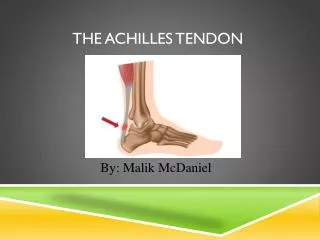
Achilles Tendon Is the large tendon formed by the medial and lateral gastrocnemius muscles and the soleal muscle, inserting onto the Calcaneum. The Achilles tendon provides the power to ‘push off’ when walking or running. Achilles tendon long and trans-normal ultrasound appearance
Two of the more commonly encountered pathologies Tendinosis and Paratendinitis symptoms and appearances compared.
Demonstrating tendinosis of the Achilles Tendon and a swollen Retro Achilles bursa Retro Achilles bursitis
Haglund’s Deformity possible cause of Retroachilles swelling/burstis Haglund’s deformity bursitis Fig 3(3) Medical multimedia group
Retrocalcaneal Bursa. Situated between the Achilles tendon and the Calcaneum- pain similar to the presentation of Achilles tendinosis Retrocalcaneal bursitis
Increased vascularity within Achilles tendon and the retrocalcaneal bursa. Increased vascularity –paratenon peritendinitis Increased vascularity within the Achilles tendon and retroachilles bursa Note:- always relax the foot when Using colour so as not to compress the vessels.
An intact Plantaris tendon can cause a false impression of a partially torn Achilles tendon. Care must be taken to assess the Plantaris Tendon and to be aware that when performing Thompsons’ manoeuver- squeezing the calf to produce plantar flexion of the foot which indicates an intact Achilles Tendon that the Plantaris tendon may cause plantar flexion of the foot in a ruptured Achilles, leading to a false impression of intact Achilles tendon.
References 1. Muscle system pro.III 3d4Medical. Ipad Application.2012 available from www.Apple.com/app 2. Netta. Elsiever. Ipad Application. 2011.available from www.Apple.com/App 3. Medical Multimedia group www.orthi=ogate.org/patient-education/foot