Download
1 / 30
520 likes | 1.66k Views
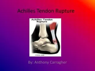
Achilles Tendon Rupture. M.Mazloumi MD. Anatomy. Largest tendon in the body Origin from gastrocnemius and soleus muscles Insertion on calcaneal tuberosity. Anatomy. Lacks a true synovial sheath Paratenon has visceral and parietal layers Allows for 1.5cm of tendon glide. Anatomy.

E N D
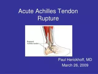
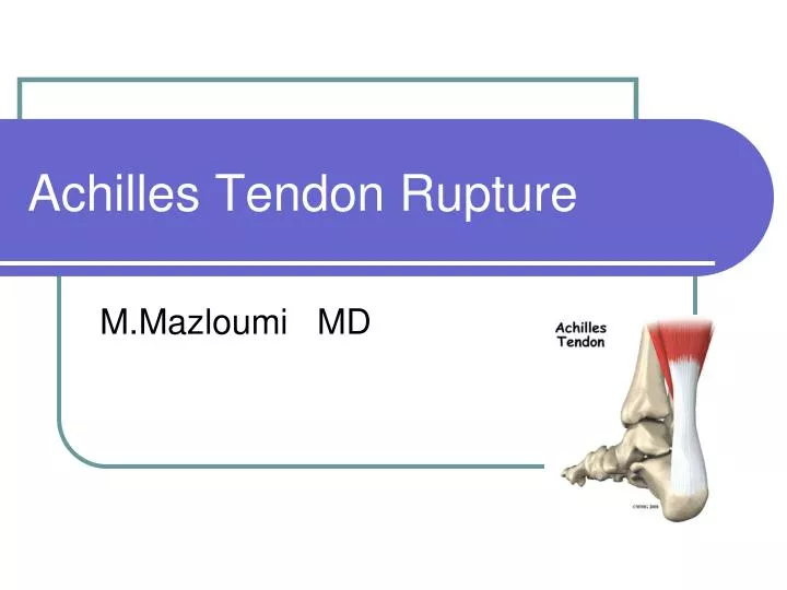
Achilles Tendon Rupture M.Mazloumi MD
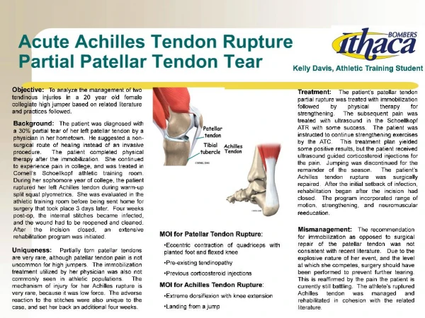
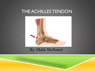
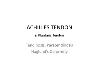
Anatomy • Largest tendon in the body • Origin from gastrocnemius and soleus muscles • Insertion on calcaneal tuberosity
Anatomy • Lacks a true synovial sheath • Paratenon has visceral and parietal layers • Allows for 1.5cm of tendon glide
Anatomy • Paratenon • Anterior – richly vascularized • The remainder – multiple thin membranes
Anatomy • Blood supply • Musculotendinous junction • Osseous insertion on calcaneus • Multiple mesotenal vessels on anterior surface of paratenon (in adipose) • Anterior mesentery • Hypovascular area at 2 to 6 cm proximal to osseous insertion
Physiology • Remarkable response to stress • Exercise induces tendon diameter increase • Inactivity or immobilization causes rapid atrophy • Age-related decreases in cell density, collagen fibril diameter and density • Older athletes have higher injury susceptibility
Biomechanics • Gastrocnemius-soleus-Achilles complex • Spans 3 joints • Flex knee • Plantar flex tibiotalar joint • Supinate subtalar joint • Up to 10 times body weight through tendon when running
Achilles Tendon Rupture • Pathophysiology • Repetitive microtrauma in a relatively hypovascular area. • Reparative process unable to keep up
Achilles Tendon Rupture May be on the background of a degenerative tendon
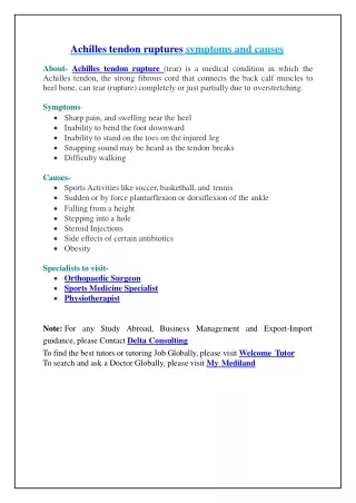
Achilles Tendon Rupture • Antecedent tendinitis/tendinosis in 11% • 75% of sports-related ruptures happen in patients between 30-40 years of age. • Most ruptures occur in 4cm proximal to the calcaneal insertion.
Achilles Tendon Rupture • History • Case reports of fluoroquinolone use, steroid injections • Mechanism • Eccentric loading (running backwards in tennis) • Sudden unexpected dorsiflexion of ankle • Direct blow or laceration • Fall from a hight
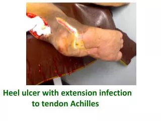
Achilles Tendon Rupture • Physical • Partial • Localized tenderness +/- nodularity • Complete • Defect • Can not heel raise • Positive Thompson test
Imaging • Ultrasound • Inexpensive , dynamic examination possible • Good screening test for complete rupture
Imaging • MRI • Expensive • Better at detecting 1-partial ruptures 2- staging degenerative changes 3- monitor healing
Management Goals • Restore musculotendinous length and tension. • Optimize gastro-soleous strength and function • Avoid ankle stiffness
Conservative Management CAM Walker or cast with plantarflexion q 2 wks Cast in Plantarflexion 2 wks 4 weeks Allow progressive weight-bearing in removable cast Start physio for ROM exercises When WBAT and foot is plantigrade 2- 4 weeks Start a strengthening program Remove cast and walk with shoe lift. Start with 2cm x 1 month, then 1cm x1 month then D/C
Surgical Management • Preserve anterior paratenon blood supply • Beware of sural nerve • Debride and approximate tendon ends • Use 2-4 stranded locked suture technique • May augment with absorbable suture • Close paratenon separately
Surgical Management Kerachow suture technique Dynamic loop suture of Peroneus brevis
Surgical Management Lynn technique Percutaneous repaire
Old rupture Bosworth technique for repairing old ruptures of Achilles tendon Wapner technique with FHL tendon
Percutaneous versus open repair Percutaneous repair Open repair
Surgical Management : Post– op Care • Assess strength of repair, tension and ROM intra-op. • Apply cast with ankle in the least amount of plantarflexion that can be safely attained. • Patient returns to fracture clinic 2 weeks post-op.
Conservative vs Surgical Acute rupture of tendon Achillis. A prospective randomised study of comparison between surgical and non-surgical treatment.Moller M, et al. J Bone Joint Surg Br. 2001 Aug;83(5):863-8 112 patients Casted x 8 wks Surgery + Early functional rehab in brace 21 % re-rupture 1.7% re-rupture 5% infection 2% Sural nerve inj. No difference in functional outcome
Conservative vs Surgical Acute Achilles tendon rupture: minimally invasive surgery versus nonoperative treatment with immediate full weightbearing--a randomized controlled trial. Am J Sports Med. 2008 Sep;36(9):1688-94. Epub 2008 Jul 21. 83 patients Casted x 8 wks Surgery + Early functional rehab in brace 5 \ 41 re-rupture 3 \ 42 re-rupture 0.5% infection 0.1% Sural nerve in No difference in functional outcome
Outcome of achilles tendon ruptures treated by a limited open technique. Jung HG, Lee KB, Cho SG, Yoon Foot Ankle Int. 2008 Aug;29(8):803-7. Repair of achilles tendon rupture under endoscopic control. Fortis AP, Dimas A, Lam Arthroscopy. 2008 Jun;24(6):683-8. Minimally invasive repair of ruptured Achilles tendon. Chan SK, Chu Hong Kong Med J. 2008 Aug;14(4):255-8. Limited open technique