Download
1 / 42
420 likes | 629 Views
THE MULTIPLE ROLES OF COMPLEMENT. Dr Andrew Guirguis Haematology Registrar The Alfred Hospital Scientific Meeting – 22 nd May, 2008. History of complement. Ehrlich – role of ‘complementing’ antibodies in killing of bacteria. 1895 – Bordet Subsequent discovery of components

E N D
THE MULTIPLE ROLES OF COMPLEMENT Dr Andrew Guirguis Haematology Registrar The Alfred Hospital Scientific Meeting – 22nd May, 2008
History of complement • Ehrlich – role of ‘complementing’ antibodies in killing of bacteria. • 1895 – Bordet • Subsequent discovery of components • Current knowledge:- • > 30 proteins in plasma + on cell surfaces • ~ 15% of globulin fraction of proteins
Nomenclature • C1 – C1q, C1r, C1s • C4, C2, C3, C5, C6, C7, C8, C9 • Many referred to as ‘zymogens’ • ‘a’ and ‘b’ – added in to denote cleavage products. • ‘b’ – larger fragment • Alternative pathway proteins:- ‘Factors’ or identified by single letters • Complement receptors:- named according to ligand (eg C6 receptor) or using CD system.
The basics! • ‘Innate immune system’ • Cascade • C3 – most important component • Activation:- innate or adaptive systems • Classical:- adaptive immune system – immune complexes bind to C1q • Alternative:- innate – chance binding of C3b to microorganism surface. • Distinction of self from non-self! • Deficiencies:- increased susceptibility to recurrent infections (pyogenic bacteria) OR illnesses a/w production of autoantibodies + immune complexes.
Main roles • Defends against pyogenic bacterial infections • Bridges both the innate and adaptive immunity systems • Assists in disposing of immune complexes etc
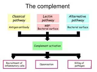
Role in Inflammation • Opsonization:- C3b is important! • Chemotaxis:- complement fragments diffuse from target – stimulating cellular movement and activation. • Target cell lysis:-‘membrane attack complex hydrophobic ‘plug’ inserted into lipid membrane bilayer
Activation • Pathways:- 1. Classical 2. Lectin 3. Alternative • Common end point: formation of C3 convertase – cleaves to C3a and C3b • Classical + Lectin pathways – C4b2a • Alternative pathway – C3bBb • Ultimately:- converted into C5 convertase – by further addition of C3b. Production of MAC.
1. Classical pathway • ‘Antibody’ directed • Begins with C1 • C1 • Pentamolecule – C1q fragment (6 domains) + 2 x C1r + 2 x C1s • Antibody binds to two or more of the six domains (binds either IgG or IgM molecules) • C1 complex undergoes conformational change • ‘Autocatalysis’ of C1r • C1s activation
2. Lectin pathway • Antibody independent • C1q – calcium-dependent lectin (collectin) • Other members:- mannan-binding lectin (MBL), conglutinin and lung surfactant A + D. • MBL – may bind mannose grps on bacterial surface – then interacts with associated Serine Proteases – MASP1 and 2 (homologous to C1r and C1s). • Antibody independent activation of classical pathway
Downstream effects • C1 – cleaves C4 – forming activated C4b • Two isotypes exist • C4A – binding amine grps (usually on proteins) • C4B – hydroxyl grps on CHO • C4b – allows binding of C2. Acted on by C1s to release C2b. • C4b + C2a = classical pathway convertase (C3) • By definition:- C3 convertase – breaks up C3 to C3a and C3b (focus of further complement activation)
What about regulation? • C1 inhibitor – serine proteinase inhibitor (aka serprin) – binds and inactivates C1r and C1s • Inhibition of formation of C3 convertase enzyme- C4b2a (by ongoing catabolization of C4b by Factor I and C4 binding protein) • Other complement control factors – inhibit complement binding to host cell surfaces • DAF: (Decay accelerating factor) – CD55 • CR1 • MCP: Membrane co-factor protein • Inhibit binding of C2 to C4b; promote ‘decay acceleration’ of C2a from C4b. Assist in catabolism of C4b by Factor I
Alternative pathway • Spontaneous activation – C3 is susceptible to spontaneous hydrolysis by water • ‘Tick over activation’ – to form C3i • C3i – acts as binding site for Factor B (cleaved by Factor D – to form Ba) • C3iBb – alternative pathway C3 convertase • Most C3b generated becomes inactivated in water. If it comes into contact with non-self – initiates amplification loop of alternative pathway.
Regulation… it’s always about rules!!! • Factor H and I • DAF + CR1 – accelerate dissociation of C3bBb ‘How C3b reacts is governed by the surface to which it attaches’ – protected vs non-protected
Initiators of complement activation pathways • Classical • Immune complexes • Apoptotic cells • Viruses + GN bacteria • CRP bound to ligand • Lectin • Mannose groups – terminal ends of microbes • Alternative • Bacteria, fungi, viruses, tumour cells etc
Membrane attack complex • Requires enzymatic cleavage of C5 • Sequential binding of C6, C7 (hydrophobic status), C8, C9 (up to 14 monomers) • Formation of lytic ‘plug’ – majority of damage caused by C9 • C9 – analogous to perforin (used by T lymphocytes) • C5b67 – can be inactivated by numerous means (S protein – vitronectin etc) • RBC immunity: poorly lysed by homologous complement • CD59: glycophospholipid foot. Inhibits insertion + unfolding of C9 into membranes.
Clinically speaking… • CH50 / THC (total haemolytic complement):- requires all nine components of classical pathway to give normal value – used to screen for deficiency of classical pathway. • If very low - ? Homozygous deficiency of classical pathway component • Less dramatic reduction during inflammatory process • AH50: alternative pathway measure • C3/4:- helpful as activity markers in those with SLE • Anaphylatoxins:- C5a / C3a – if increased – complement activation • ? M’ment of split products
Elevated complement levels = inflammatory response (i.e acute phase reaction) – esp C3 / C4 / B • Reduced levels: often a/w disease involving immune complexes / autoantibodies. May be useful for Dx + Mx of certain diseases (eg SLE, Sjogren’s, vasculitis etc) • Low C4 / C3 + N FB – classical pathway activation • Low FB + C3 + N C4 – alternative p’way activation • C4 + FB – low = both p’ways activated
Clinical implications • Complement deficiencies • Glomerulonephritis • C1 inhibitor deficiency • SLE • PNH • Sepsis • APLS
1. Complement deficiency:-Increased susceptibility to pyogenic infections • Contributing factors • Deficient opsonisation • Deficiency compromising lytic activity • Deficient manose-binding lectin pathway • Pyogenic infection:- • Site of defect:- antibody production, complement proteins of classical pathway, phagocyte function • Usually bacteria is opsonised with Ab – complement is then activated, phagocytosis occurs and intracellular killing • Key player:- C3b • Impaired lysis • MAC component deficiency – a/w Neisserial disease* • Risk of meningococcal disease ~ 0.5% / yr (RR 5000 cf normal population) • Deficient lectin • Deficiency occurs due to 1 of 3 point mutations – a/w reduced levels. • Associated with higher risk of infection in children – whilst losing passive immunity • ? Protective against mycobacterial infections
2. Glomerulonephritis… Key of C3b regulation:- whether Factor B or H binds to C3b • If C3 regulation is defective:- often a/w GN. • Due to C3 nephritic factor – increases stability of C3 convertase enzymes – association with membranoproliferative GN OR • Reduced function of Factor H or I • ? Associations with HUS (+/- low level of C3)
3. C1 INHIBITOR DEFICIENCY • Autosomal dominant – inadequate production of physiologically adequate C1 inhibitor • Type 1:- 85% - reduced transcription of abnormal allele. Reduced levels of C1 inhibitor • Type 2:- point mutation in C1 inhibitor gene – altered activity (So levels may be normal or high – as not consumed) • Autoantibodies against C1 inhibitor • Inhibits – C1r and C1s, activated FXI and XII • Consumed by plasmin – trigger for angioedema attacks. • Rx: C1 inhibitor infusion.
4. Complement deficiency + SLE • Inverse correlation with position of deficient protein in activation sequence of the classical pathway • Homozygous def of C1q, C1r and C1s + C4 – strongly a/w SLE (93%, 57%, 75%) • Cf. def of C2 – 10% prevalence. • Protective role exists for those in whom activation of classical pathway up to C4 cleavage occurs.
5. PNH:- • Acquired stem cell disorder • Deficiency of PIG-A (somatic mutation) – required for synthesis of glycosyl-PI phospholipid. • Important for anchorage of proteins to cell membranes • In PNH – lack of GPI-linked proteins (including complement-regulating surface proteins) - eg DAF (i.e CD55) which regulates formation of C3 convertase and CD 59 – restricts formation of MAC. • Deficiency on RBCs:- does not allow protection against terminal complement • Clinically: chronic haemolysis, fatigue, pain, thrombotic events. median age – early 30s; median survival as low as 10-15 yrs.
Smooth muscle dystonia - ? 2’ to NO depletion during chronic haemolysis
Who to screen? • Hb’uria • Coombs –ve haemolytic anaemia • Those with AA or MDS (annual screen) • Haemolytic anaemia • VT without explanation (including unusual sites – eg mesenteric, portal, cerebral etc) • Unexplained arterial thrombosis • Episodic dysphagia or abdo pain Parker et al, 2005
Dx:- • Flow cytometry: gold standard (peripheral blood). Granulocytes provide best estimate of PNH clone size.
Role of Soliris (eculizumab) • Other Rx:- • Supportive transfusions • Haematinic supplementation • Anticoagulation (for those with Hx of thrombosis or for prophylaxis) • Therefore multiple benefits • Risks??
6. Complement system + sepsis • C5a – anaphylatoxin – strong chemoattractant. • Sepsis – excessive early production of C5a – upregulated proinflammatory response. • ? Role for blockade of C5a with antibodies – shown to improve survival of septic mice. • ? Use in IHD to assist cardiac reperfusion
7. Complement + APLS • Nature medicine 2004:- • Previous mouse models – shown that complement activation plays an important role in pregnancy + fetal growth restriction • Likely induced by activation thru aPL antibodies (classical pathway) • Anticoagulation alone – insufficient in completely averting miscarriage • Heparin use - ? Additional role via inhibition of complement.
APLS • Mouse model used:- • Pregnant mice injected with aPL antibodies • Rx:- heparin (UFH or LMWH) – reduced frequency of fetal resorption to that of healthy controls. • To rule out mere ‘anticoagulant effect’ – use of fondaparinux or hirudin – both do not directly affect the complement systems.
In vivo:- • Focus on C3 and degradation products – increased levels seen with aPL-IgG injection. • Abolished by UFH or LMWH, but not by fondaparinux or hirudin. • Separate study:- use of Crry-Ig (complement receptor 1-related gene / protein y)(exogenous inhibitor of C3 activation) OR C3 deficient mice – similar results. • Associated with fewer resorptions and less antibody-mediated growth retardation • Activation of complement – associated with thrombophilic state