Download
1 / 11
311 likes | 2.28k Views
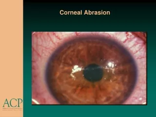
CORNEAL DEGENERATIONS. 1. Age-related. Arcus senilis. Vogt white limbal girdle. Crocodile shagreen . Cornea guttata. 2. Lipid keratopathy. Primary. Secondary. 3. Band keratopathy. 4. Spheroidal degeneration. 5. Salzmann nodular degeneration. Arcus senilis.

E N D
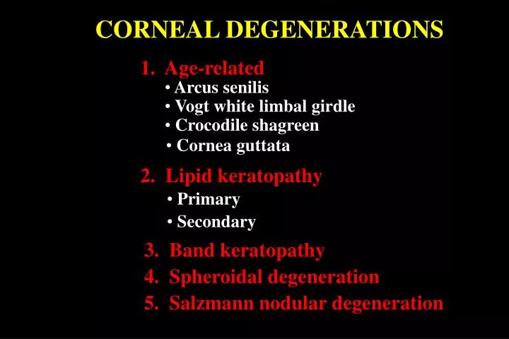
CORNEAL DEGENERATIONS 1. Age-related • Arcus senilis • Vogt white limbal girdle • Crocodile shagreen • Cornea guttata 2. Lipid keratopathy • Primary • Secondary 3. Band keratopathy 4. Spheroidal degeneration 5. Salzmann nodular degeneration
Arcus senilis • Innocuous and extremely common in elderly • Occasionally associated with hyperlipoproteinaemia • Peripheral border separated • from limbus by clear zone • Bilateral, circumferential bands • of lipid deposits • Diffuse central and sharp • peripheral border • Clear zone may be thinned • ( senile furrow)
Vogt white limbal girdle • Innocuous and very common in elderly • Bilateral • White, crescentic line along nasal and temporal • limbus • Type 1 - separated from limbus by clear zone • Type 2 - not separated by clear zone
Crocodile shagreen • Uncommon and innocuous • Usually bilateral • Polygonal stromal opacities separated by • clear space • Most frequently involve anterior stroma • (anterior crocodile shagreen) • Occasionally involve posterior stroma • (posterior crocodile shagreen)
Cornea guttata • Common, bilateral and usually innocuous • Rarely progression to Fuchs dystrophy • Tiny dark spots on central endothelium • Similar peripheral lesions are Hassell-Henle bodies
Lipid keratopathy Primary Secondary • Common, secondary to previous • disciform keratitis • Rare, occurs spontaneously in • avascular cornea • Usually unilateral stromal deposits • without vascularization • Unilateral stromal deposits with • vascularization Treatment - coagulation of feeder vessels and/or keratoplasty Treatment - keratoplasty, if severe
Band keratopathy • Common, unilateral or bilateral depending on cause • Subepithelial calcification Progression • Central spread of calcification • Interpalpebral limbal opacification • Small holes within calcified area • Separated by clear zone
Causes of Band Keratopathy 1. Ocular (common) • Chronic iridocyclitis, particularly in children • Associated with phthisis bulbi • Silicone oil in anterior chamber 2. Metabolic (rare) • Increased serum calcium and phosphorus • Hyperuricaemia • Chronic renal failure 3. Hereditary (rare) • Familial band keratopathy • Hereditary ichthyosis 4. Age-related (uncommon)
Chelation of band keratopathy Application of sodium versenate Removal of corneal epithelium
Spheroidal degeneration • Rare, typically affects outdoor workers • Starts with peripheral, interpalpebral, small amber-coloured • granules in superficial stroma Progression Central spread, coalescence and opacification Advanced lesions become nodular and elevated Treatment • Debridement or superficial keratotomy if mild • Keratoplasty if severe
Salzmann nodular degeneration • Uncommon, unilateral or bilateral • Secondary to chronic keratitis • Discrete superficial stromal opacities • and nodules • Base of nodule may be surrounded by • iron deposits Treatment- similar to spheroid degeneration