Download
1 / 32
320 likes | 1.21k Views
Case Presentation. 62 year old male with 8 month history of increasing hoarseness. Over the past 3 days he has had increased difficulty breathing and swallowing. He comes to the emergency room for evaluation because he

E N D
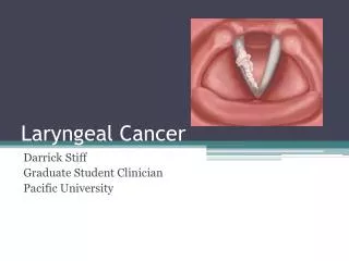
1. Laryngeal Mass John F. McGuire, MD, MBA
2. Case Presentation 62 year old male with 8 month history of increasing hoarseness. Over the past 3 days he has had increased difficulty breathing and swallowing. He comes to the emergency room for evaluation because he �cant breathe�.
3. History PMHx: none
PSHx: none
Meds: none
PHx: 40 pack year smoking history, weekly to daily alcohol intake
4. Exam Pt has biphasic stridor and appears in distress.
Next step???
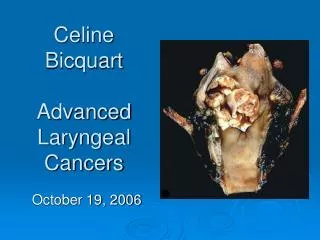
Findings in OR: �Exophytic obstructive transglottic lesion encompassing the left supraglottic, left glottic and small extention into the left subglottic regions.�
6. Differential Dx
V: hemangioma
I: TB (20-40% with no lung involvement, used to be MC disease affecting larynx), histoplasmosis, blasto/coccidio/actino-mycosis, cryptococcosis, Feinberg�s sequence
T: trauma, fb
A: relapsing polychondritis, rheumatoid arthritis (MC autoimmune dz to affect larynx, laryngeal involvement in 26% to 53%), and Wegener's granulomatosis (23% with laryngeal involvement� think subglottic stenosis: tx??? A: cut, dilate, mitomycin� not laser)
M:
I: sarcoidosis, amyloidosis
N: granular cell tumors (MC non-epitheliod lesion of larynx), chondrosarcoma, fibrosarcoma, kaposi�s sarcoma, adenocarcinoma, mucoepidermoid carcinoma, atypical carcinoid (most common neuroendocrine tumor of larynx), extramedullary plasmacytoma
C:
Common things common: 95% of larygeal CA is SCCA
7. Topic of this Presentation Optical coherence tomography of the vocal cords
OCT is like ultrasound, but its �light� instead of �sound��
think of it as �ultralight��
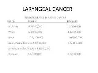
8. Laryngeal Cancer Hypopharynx cancers: Glottic (62%) supraglottic (37%) and subglottic (2%).
Main risk factors: tobacco and alcohol
Others include HPV, GERD, passive smoke, (for the test� Plummer-Vinsen Syndrome >> post-cricoid SCCA)
9. Clinical Pearls 78% of patients with hypopharyngeal carcinoma have palpable cervical metastases when initially seen.
The average duration of symptoms before presentation is 2 to 4 months.
20% have an asymptomatic mass in the neck, usually ipsilateral, a jugulodigastric or midjugular lymph node
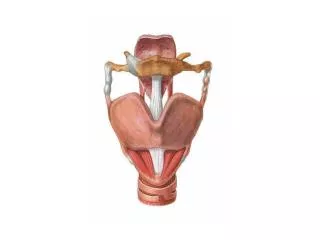
10. Anatomy: Think Spaces Quadrangular membrane: Fibrous drape from epiglottis over arytenoids.
Conus elasticus: See diagram.
Anterior commissure tendon (Broyles ligament):
- No perichondrium.
Hyoepiglottic ligament:
Roof of the paraglottic and preepiglottic spaces.
11. Anatomy Paraglottic space:
Superior border : quadrangular membrane
Inferior border: conus elasticus
Lateral border: inner surface of the thyroid cartilage
Medial border: ventricle
13. Anatomy Preepiglottic space
Superior border : hyoepiglottic ligament
Anterior border: thyrohyoid membrane and ligament
Posterior border: anterior surface of the epiglottis and thyroepiglottic ligament
Clinical note: Almost 50% of supraglottic carcinomas have preepiglottic space involvement� implication is upstage to T3 tumor. This is likely secondary to fenestrations in epiglottic cartilege.
15. Anatomy and Cancer Weak points for the spread of laryngeal cancer
Broyle�s ligament has no perichondrium, providing carcinoma direct access to the cartilage.
Fenestrations within the infrahyoid epiglottis provide a route for invasion of the preepiglottic space.
Ossification at the anterior commissure and the posterior border of the thyroid ala of the thyroid cartilage provide a route for cancer spread.
Points of attachment of the cricothyroid ligament and the anterior origin of the thyroarytenoid musculature provide a route for cancer spread.
16. Anatomy: Lymph Drainage Rule of thumb: Glottic and supraglottic to levels 2-3, subglottic to level 4
Very sparce lymphatics in TVC, therefore glottic CA usually better prognosis (although also usually detected earlier)
Delphian node = midline pretracheal node
Clinical notes:
Correlation of presumptive nodal abnormality on physical exam with pathologic study has been 60% to 70% and varies between approximately 65% and 80% for imaging studies.
Glottic and subglottic tumors have a 2% to 5% risk of neck disease unless the subglottic extension exceeds 10 mm.
17. Radiology Only 46% positive predictive value of CT for detecting cartilage invasion
50% of tumors radiologically staged as T3 had microinvasion of cartilage, usually at thyroid notch.
CT most useful, but MRI useful for detecting submucosal lesions.
Don�t forget CXR: lung CA most common second primary.
18. Staging: Glottic T1-tumor limited to the vocal cords
T1a-tumor limited to one vocal cord
T1b-tumor involves both vocal cords
T2-tumor extends to the supraglottis and/or subglottis and/or impaired vocal cord mobility
T3-tumor with vocal cord fixation (a cord cannot move at all)
T4-tumor invades outside of the larynx (trachea, soft tissues of the neck, etc.)
19. Staging: Supraglottic T1-tumor limited to one subsite of the supraglottis with normal vocal cord mobility
T2-tumor invades one adjacent site of the supraglottis or glottis or one region outside of the supraglottis without fixation of the vocal cords
T3-tumor limited to the larynx with vocal cord fixation or invasion into the area behind the larynx or in front of the larynx
T4- tumor invades outside of the larynx (trachea, soft tissues of the neck, etc.)
20. Staging: Nodal Disease N0-no spread to lymph nodes
N1-tumor spread to one lymph node on the same side as the tumor in the throat. Lymph node must be < 3 cm
N2a-tumor spread to one lymph node on the same side as the tumor in the throat. Lymph node is between 3 and 6 cm
N2b-tumor spread to more than one lymph nodes on the same side as the tumor in the throat, with none being >6 cm
N2c-tumor spread to lymph nodes on both sides of the neck, with none being >6 cm
N3-tumor spread to a lymph node when the lymph node is > 6 cm
21. Overall Stage Stage I-T1N0M0
Stage II-T2N0M0
Stage III-T1-3N1M0 or T3N0M0
Stage IVA-T4N0-1M0 or Any T, N2M0
Stage IVB-Any T, N3M0
Stage IV-any M1
22. Carcinoma in situ Equal efficacy rates of surgical stripping and XRT>> what to you do? Why? (10% failure with XRT in CIS)
Progression: hyperkeratosis with atypia >> CIS >> superficially invasive CA >> invasive CA
5-30% with pre-malignant lesions will develop invasive disease
Appx. 90% cure rate with stripping:
Caveats:
May not be true for anterior commisure lesions
Can require re-stripping
Need reliable patient, second look 6-12 weeks out standard
23. Organ Sparing Surgery Principles:
Local control and accurate assesment of 3D extent of tumor
The cricoarytenoid unit is the basic functional unit of the larynx.
�It is the cricoarytenoid unit, not the vocal folds, that allows for physiologic speech and swallowing without the permanent need for a tracheostoma after supracricoid laryngectomy.�
Resection of normal tissue to achieve consistent functional outcomes in terms of speech and swallowing.
Standard resections lead to consistent functional outcomes
24. Organ Sparing Surgery Mostly for early laryngeal cancers (T1 and T2)
Absolute Contraindications:
arytenoid fixation, thyroid cartilage invasion, interarytenoid invasion, subglottic extension to involve the cricoid cartilage, lesions that extend outside the larynx, and preepiglottic space invasion.
(a relative contraindication is anterior commisure lesions� recurrance rates are higher and speech results are variable)
Preoperative evaluation
�fixed vs. pseudofixed� TVC
Pulmonary function testing:
the real issue is how well pt will tolerate aspiration in early recovery period
COPD is relative contraindication
25. Vertical Hemilaryngectomy
26. Supracricoid Partial Laryngectomy (SCLP) All you can eat: resects of both true cords, both false cords, the entire thyroid cartilage, both paraglottic spaces bilaterally, and a maximum of one arytenoid.
Useful for T2 and T3 lesions.
Consistently low local recurrence rates likely 2nd to the complete resection of the entire thyroid cartilage and the bilateral en bloc resection of the paraglottic spaces
Performed in conjunction with cricohyoidoepiglottopexy.
�Chronic and inefficient cough, purulent sputum, and/or an inability to climb 2 flights of stairs without shortness of breath are strong contraindications against the use of SCPL-CHEP or SCPL-CHP.�
27. SCPL
28. Transoral Laser Resection At this point, performed only by the pioneers of our specialty�
Dr. Armstrong, a few words about basic principles of the transoral laser approach???
Some have compared this approach to Moh�s surgery, would you agree?
29. Laryngeal Preservation Idea: prevent surgery with chemotherapy
Current best results from Radiation Therapy Oncology Group (RTOG), showing that concurrent cisplatin (rather than induction with cisplatin and 5-FU in the famous VA study) chemo/XRT has 88% laryngeal preservation rate.
Distant mets and localregional control best in concurrent group, but no difference in survival among groups.
Caveats:
Larygeal preservation does not imply functional larynx >> chondronecrosis and aspiration are complications >> up to 40% of �preserved� larynges require laryngectomy
Frequent problems with dysphagia/strictures
Rate of fistula after faile laryngeal preservation goes up to 30-60%
30. Neck Dissection in No Neck getting XRT? Incidence of occult mets varies by site, with glottic being lowest (15%), supraglottic more (20-38%), and piriform sinus at highest.
Any N should get ND.
31. Complications of TL Early Complications
Drain failure, Hematoma., Infection, Chyle fistula
Wound dehiscence: Local wound care should suffice for healing by secondary intention, but if the carotid becomes persistently exposed, vascularized muscle flap coverage is advisable.
Pharyngocutaneous fistula.
At risk: poor nutritional status, positive surgical margins, preoperative radiotherapy.
Such fistulas may occur 1 to 6 weeks postoperatively,
confirmed by a methylene blue swallowing test.
Management: fistula-track packing, dressings, antibiotic therapy, NPO, diversion, pressure dressing;operative closure should be considered after 2-3 weeks.
Late Complications
-Stomal stenosis, Pharyngoesophageal stenosis and stricture, Chronic pharyngocutaneous fistula. Hypothyroidism.
32. Chyle Fistula Chyle fistula 1600 cc/24 hrs. Tx?
A: If chyle exceeds 600 mL >> early surgical exploration
prevents adherent fibrinous material and inflammation.
B: If Chylous fistulae becomes apparent only after enteral feedings are resumed, and particularly those that drain less than 600 mL of chyle per day
>>> conservative: closed wound drainage, pressure dressings, and low-fat nutritional support.
C: General:
the area should be observed for 20 or 30 seconds while the anesthesiologist increases the intrathoracic pressure.
Even the smallest leak of chylous material should be pursued seriously until it is arrested. Direct clamping and ligating may be difficult and sometimes counterproductive because of the fragility of the lymphatic vessels and the surrounding fatty tissue.
Hemoclips are ideal to control a source of leakage that is clearly visualized.
33. Stomal Recurrence Prevention of stomal recurrence in tumor with one cm transglottic spread.
postoperative stomal XRT.
paratracheal lymph node dissection.
Stomal recurrence classified by Sisson:
Type I � tumor involves the superior one half of the stoma without esophageal involvement.
Type II � tumor involves superior one half of the stoma with esophageal involvement, or the inferior half of the stoma
Type III � Tumor involves the inferior one half of the stoma and extends into the mediastinum.
Type IV-Extends beneath the clavicle