Download
1 / 32
570 likes | 1.13k Views
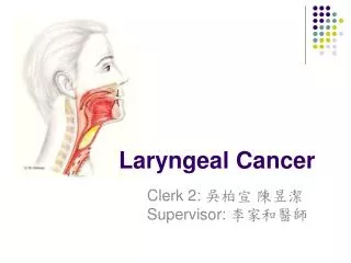
Laryngeal Trauma. Introduction. Incidence: 1:30,000 emergency patients Airway Voice Outcome determined by initial management. Anatomy and Physiology of Larynx. Well protected (mandible, sternum, neck flex) Functions: Airway, tracheobronchial protection, voice

E N D
Introduction • Incidence: 1:30,000 emergency patients • Airway • Voice • Outcome determined by initial management
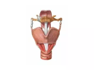
Anatomy and Physiology of Larynx • Well protected (mandible, sternum, neck flex) • Functions: Airway, tracheobronchial protection, voice • Support: Hyoid, thyroid, cricoid • Innervation: RLN, SLN • Supraglottis: soft tissue • Glottis: relies on external support, crico-arytenoid mobility and neuromuscular input • Subglottis: cricoid, narrowest in infants
Mechanism of Injury • Blunt – • motor vehicle accident , strangulation, clothesline, sports related • Significant internal damage, minimal external signs • Penetrating • Gun shot wound: damage related to velocity • Knife: easy to underestimate damage
Blunt Trauma: Mechanisms of Injury • Compression over spine • Static lateral force • Laryngo-Tracheal separation
Initial Evaluation • Secure airway – local tracheotomy • Intubation can worsen airway • Avoid cricothyroidotomy • Pediatric: tracheotomy over bronchoscope
History • Change in voice – most reliable • Dysphagia • Odynophagia • Difficulty breathing - more severe injury • Anterior neck pain • Inability to tolerate supine position – probable airway compromise imminent
Physical exam • Stridor • Hoarseness • Subcutaneous emphysema • Hemoptysis • Laryngeal tenderness, ecchymosis, edema • Loss of thyroid cartilage prominence • Associated injuries - vascular, cervical spine, esophageal
Flexible Fiberoptic Laryngoscopy • Perform in emergency room • Findings dictate next step • CT scan • Tracheotomy • Endoscopic • Surgical Exploration • Other studies
Radiographic Imaging • C-spine • CT if airway stable and mild abnormality on flexible exam. • Good for intermediate cases with scope limited by edema • Angiography and contrast esophagrams considered
CT Scan Indications: • Significant mechanism of injury • Rule out occult fracture/dislocation • Confirmation of suspectedfracture • Determine extent of fracture(s)
Laryngotracheal Injury Classification • Group I: Minor hematoma, no fracture • Group II: Edema/hematoma, minor mucosal injury, no exposed cartilage, non displaced fracture • Group III: Massive edema, mucosal tears, exposed cartilage, cord immobility • Group IV: See group III, more than 2 fracture lines, massive trauma laryngeal mucosa • Group V: Complete laryngotracheal separation (Schaefer, 1982)
Laryngeal Trauma Asymptomatic or minimal symptoms F/L CT scan Displaced fracture (by CT or exam) Loss of mucosa or extensive laceration Bleeding Exposed cartilage Mild Edema Small hematoma Non-displaced linear fracture Intact mucosa Small lacerations Tracheotomy Bed rest Cool mist Antibiotics Steroids Anti-reflux Panendoscopy Explore
Laryngeal Trauma Respiratory distress, open wounds, bleeding Tracheotomy Panendoscopy Explore
Comminuted fractures Displaced fractures All fractures involving the median and paramedian thyroid ala Cricoid fracture LT separation Large mucosal lacerations Laceration of AC and free edge VC Disruption CA joint VC immobility Exposed cartilage Indications for Repair
Goals of Laryngeal exploration • Cover all cartilage to prevent granulation tissue and fibrosis • Primary closure ideal,can undermine mucosa or use advancement flaps from epiglottis or pyriforms • Palpate arytenoids and reposition if necessary • Resuspend anterior commisure, ORIF of fractures
Treatment Goals • Preservation of airway • Prevention of aspiration • Restoration of normal voice
Outcomes • Airway • Poor – trach dependent • Fair – mild aspiration or exercise intolerance • Good – preinjury status
Outcomes • Voice • Poor: aphonia or whisper • Fair: changed or hoarse • Good – normal voice
Outcomes • Swallowing • Normal • Abnormal • Subjective patient report
Outcomes • Medical better than surgical • Voice results worse with use of stents (airway the same), less time in better • Vocal cord paralysis – poorer outcome • Improved results with repair <48 hours
Conclusions • Rare injury • Assess airway first and follow systematic management • Timely evaluation with high index of suspicion for classic signs and symptoms • Don’t forget about associated vascular or esophageal injuries • Treatment based on site/extent of injury