Download
1 / 44
500 likes | 1.49k Views
Urinary system Systema urinarium. Anatomy + a glimpse of histology and embryology. Upper urinary tract Nephron Tubuli colligentes Lower urinary tract Calices renales (Renal calices) Pelvis renalis (Renal pelvis) Ureter Vesica urinaria ( Urinary bladder ) Urethra feminina / masculina.

E N D
Urinary systemSystema urinarium Anatomy + a glimpse of histology and embryology
Upper urinary tract • Nephron • Tubuli colligentes Lower urinary tract • Calices renales (Renal calices) • Pelvis renalis (Renal pelvis) • Ureter • Vesica urinaria (Urinary bladder ) • Urethra feminina / masculina
Kidney (Ren, Nephros) • margo, hilum, sinus, facies, extremitas /polus/ • capsula fibrosa, lobi renales • Cortex: labyrinthus, columnae renales Bertini, radii medullares • Medulla: pyramides renales → papillae renales (area cribrosa + foramina papillaria) • zona interna + externa
Kidney Microcopic structure • Nephron • Interstitium • Vessels
Nephronfunctional unit • Corpusculum renale Malphigi • glomerulus (vessel skein) • capsula glomeruli Bowmani • Proximal tubule • Intermediate tubule • Distal tubule • Juxtaglomerular apparatus
Capsula glomeruli (Bowman´s capsule) parietal lamina (flat monolayer epithelium) visceral lamina (podocytes with pedicles) Glomerulus vas afferens vas efferens (= arterioles) fenestrated capillars without diaphragm Corpusculum renale(ledvinné tělísko Malpighi) • Filtration barrier basal membrane - colagen IV. typ and heparan sulfate negative charge • Mesangial cells cleaning function of filter
Proximal tubule • single layer of cuboidal epithelium rich in microvilli • resorption of NaCl and water (80-95%) • Na+ passive transport into cells, active transport out of cells
Intermediate tubulus • = thin segment of ansa nephroni (Henle´s renal loop) • descending and ascending limb • flat cells, few organels • descending segment permeable for water • ascending not!! • juxtamedullary nephrons with long H. loop • countercurrent together with vasa recta
Distal tubule • cuboid eptihrelium with few microvilli • back-resorption Na+ and secretion K- • macula densa - chemoreceptors (Cl-) • includes the thick segment of Henle´s loop
Juxtaglomerullar apparatus • components • macula densa of distal tubulus • granullar cells (transformed smooth muscle cells) of vas afferens + efferens • mesangial cells • function • regulation of blood pressure by hormone renin
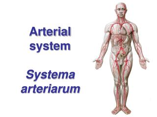
Kidney - arterial supply I. • a. renalis (paired visceral branch of abdominal aorta at level of discus intervertebralis L1/2, left L1) • accessory renal artery (30%) • rate flow 1,2-1,3 l blood/min • arteries are terminated = no arterio-arterial anastomosis
Kidney - arterial supply II. a. renalis r. anterior 4 segmental branches r. posterior for 1 dorsal segment aa. segmentales a. lobares (about 12) 2-3 aa. interlobulares aa. arcuatae a. interlobulares arteriolae glomerulares afferentes glomerulus of capillars arteriolae glomerulares efferentes peritubullar capillary plexus orarteriolae rectae along intermediate tubulus of juxtaglomerullary nephrons
Kidney - venous supply vv. stellatae (from surface) + venulae rectae (along intermediate tubulus of juxtaglomerullary nephrons) + peritubullar capillary plexus vv. interlobulares vv. arcuatae vv. interlobares v. renalis v. cava inferior Portal system (= rete mirabile) – 2 capillary nets one after the other migthy veno-venous anastomosis
KidneyLymph drainage and innervation • 3 plexus (peritubullar, subcapsullar and from adipose capsule) • nodi lymphoidei lumbales • plexus renalis (autonomous, viscerosensitive) from ganglion coeliacum + plexus coeliacus, from ganglion aorticorenale • from n. splanchnicus minor and plexus aorticus
Kidney examination • Native X-ray snap • Sonography • Excretory urography • Ascending pyelography • Clearence • CT, MR
Kidney diseases • Development defects – agenesis, shape deformities • - cysts • Glomerulonephritis • Pyelonephritis • Hydronephrosis • Nefrohydrosis • Diabetes mellitus – nephropathy • Tumours • Grawitz´s (solitary metastasis) • Wilms´ (autosomally heridatory - children) • Renal colic
Development of urinary system • Development of kidney • Pronephros • Mesonephros • Metanephros • Development of urinary tract
Pronephros • From intermediate mesoderm of cranial 12-13 somites • from 21st day (4 primary segments) • rudimentary, disappears quickly • common presistent ductus mesonephricusWolffi
Mesonephros • origin from nephrogenic blastema • sac elongates to ductus Wolffi • approximately from 23rd day • glomerulus and collective tubules • caudal parts are origin for genital organs • = urinary system of fish
Metanephros • definitive stadium of kidney • nephrogenic blastema 3rd to 5th lumbar somites • development begins at the end of 5th week • relative ascent during development • ureteral bud sprins upwards from ductus mesonephricus Wolffi and grows in blastema • induction of maturation of nephron ureteral bud ureter, pelvis, calices, collecting tubules metanephrogenic blastema nephron
Development malformations • Atypical shapes • Cystic, polycystic kidneys • Agenesis of kidney • Ureter duplex, fissus
Kidney transplantation • since 50s´ • 5 years of graft survival - 70% • including proximal part of ureter and its blood supply (branches of renal vessels) • placed to fossa iliaca and renal artery joined to a. iliaca ext. (end-to-side) or to terminal part of a. iliaca int. (end-to-end) • if present, a. renalis accessoria joined to a. epigastrica inferior
Lower urinary tractgeneral wall structure • Mucosa (tunica mucosa) • epithelium transitional • lamina propria mucosae (collagen) • Smooth muscle (tunica muscularis) • Adventitia (tunica adventitia)
Renal pelvis (Pelvis, pyelon)Renal calices (Calices renales) • 7-14 calices minores around papilae 2-3 calices majores ureter • ampullary / branching type (typus dendriticus) • muscular layer – strong circular • projection: processus costalis L1 • blood supply: branches of renalartery, tributaries to renal vein
Ureter • 25-30 cm, lumen 4-7 mm • 3 parts: pars abdominalis, pelvina, intramuralis • crossings of important structures • 3 constrictions – risk of stone impact obstruction hydronephrosis afunction of kidney • 2 muscular layers • inner longitudinal • outer circular • adventitia
Ureter - supply • Arteries: a. renalis, aorta abdominalis, a. testicularis/ovarica, a. ductus deferentis / uterina, a. vesicalis inf. rr. ureterici • Veins run along arteries • Lymph drainage: nll. lumbales (aortici lat.), iliaci int., ext., communes • Nerves: plexus uretericus plexus renalis, aorticus abd., hypogastricus sup., inf.
Urinary bladder (Vesica urinaria, Kystos) • description: fundus, corpus, apex, cervix, uvula • trigonum vesicae – ostia ureterum, plica interureterica, ostium urethrae internum, Bell´s bands • muscles: smooth • m. detrusor (parasympathetic) • m. trigoni vesicae • ♂m. sphincter vesicae (sympathetic) • projection: behind symphysis pubica (in child over s.p.)
Urinary bladder – wall structure • folds except trigonum vesicae • smooth muscle fibres create 3 layers: • inner plexiform • middle circular m. sphincter vesicae in male only! • outer longitudinal • upper surface covered with serosa (peritoneum) = partially intraperitoneal organ
Urinary bladder – supply • a. iliaca int. a. umbilicalis aa. vesicales sup. • a. iliaca int. a. vesicalis inf. • (a. obturatoria, a. glutea inf., a. uterina, a. vaginalis rr. vesicales) • Veins: plexus venosus vesicalis (connections to plexus venosus prostaticus/vaginalis) vv. vesicales v. iliaca int. • Lymph drainage: nodi paravesicales nodi iliaci int. et ext. nodi iliaci comunes • Nerves: plexus vesicalis plexus hypogastricus inf.
Urinary bladder - fixation • Ligaments: pubovesicale, puboprostaticum, rectovesicale, vesicouterinum • Smooth muscle: pubovescalis, rectovesicalis • Ligamentum umbilicale medianum (= chorda urachi) • Fascia vesicoumbilicalis Delbeti • Septum rectovesicale Denonvilliersi • Septum vesicovaginale • Spatium retropubicum Reztii
Urinary bladder - peritoneum • Fossae paravesicales • ♂ excavatio rectovesicalis = most caudal space of peritoneal cavity in male (in contact with top of glandula vesiculosa) ♀ exc. vesicouterina ♀ exc. rectouterina (= Douglas´space) = most caudal space of peritoneal cavity in female (in contact with top of posterior vaginal vault) – palpation and puncture in inflammation via vagina !
Urethrae • Male urethra is divided in 4 parts: • transitional epithelium in firts 2 parts • pseudostratified or stratifoed columnar epithelium in the rest • Female urethra • transitional epithelium in intramural part • squamous stratified
Urethra feminina (Female urethra) • 3 parts: pars intramuralis, pelvina, perinealis • ostium urethrae internum (trigonum vesicae) • accipiens, evacuans • ostium urethrae externum (vestibulum vaginae) • crista urethralis, lacunae, glandulae urethrales (Skenei s. Guérini), ductus paraurethrales (Schülleri) • smooth muscles, around urethra striated m. sphincter urethrae, m. compressor urethrae a m. sphincter urethrovaginalis • adventitia
Urethra feminina - supply • Arteries: paired branches of a. vesicalis inf. et a. vaginalis • Veins: plexus venosus vesicalis plexus venosus vaginalis vv. pudendae int. • Lymph drainage: nl.l. iliaci int., ext. • Nerves: plexus hypogastricus inf. plexus vesicalis, plexus uterovaginalis nn. vaginales
Development of lower urinary tract • Urinary bladder • developed from cranial part of urogenital sinus • urachus • separation of excretory urinary trct and genital ducts • Urethra ♀ developed from medial part of urogenital sinus ♂ developed from medial and caudal part of urogenital sinus and glandular plate
Urine continence in female • no smooth muscle sphincter in urinary bladder • elastic fibres + venous plexuses in urethra • striated muscle m. sphincter urethrae (externus) /S2-S4/ • so-called „periurethral muscles“ (in increased intra-abdominal pressure) • m. levator ani (S3-S4) • m. compressor urethrae, m. sphincter urethrovaginalis, m. bulbospongiosus (n. pudendus)
Urethra masculina (Male urethra) • 4 parts • see male genital system