Download

1 / 27

E N D
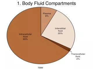
1. Cytology of Body Fluid
4. differences between transudate and exudate
5. Diagnostic role of effusion cytology It is very useful for diagnosis of premalignant and malignant tumors, especially metastatic tumors.
It is very useful for diagnosis of inflammatory conditions (septic effusion, or chronic specific inflammation e.g. TB
6. Respiratory Tract
Urinary Tract
Oral Cavity
Gastrointestinal Tract
Effusions (pleural, pericardial, joint)
Cerebral Spinal Fluid
Amniotic fluid
Many other body sites
7. Examination of body fluid Gross exam
Total cell count
Microscopic exam
Any other special test (Chemistry, Microbiology, cytology (
Test are performed in various areas of lab based on what the physician orders.
Body fluids sterile vs. non-sterile
8. Sample collection FNA of effusion fluids
Tapping
10. Fixation 1ml of heparin + 100ml of effusion fluid to prevent clotting
N.B.: do not use alcohol in fixation of fluid before spread cytological smear on glass slides
11. Types of staining smears PAP
Gram Stain
Hx & E
Cell block for remnant sediment and histopathological examination.
Other special stains for the most suspected diseases, to confirm diagnosis.
13. 1- Cerebrospinal Fluid Fluid surrounding brain and spinal cord
Sterile
Specimen collection: by Lumbar puncture
Collect 3-5 vials, each tube has a designated department.
Gross exam: Turbidity, Color, microscopic exam, cell count
14. CSF cell differential Numerate and differentiate cells seen
Lymphocytes: usually are few; increased with viral, fungal, bacterial meningitis, or nervous system disease
Monocytes: Less than 2% of normal CSF, increased with TB meningitis, viral encephalitis, subarachnoid hemorrhage.
15. PMN: are few, associated with Viral and acute bacterial inflammation.
Macrophages: are few in number associated with malignancy, hemorrhage, inflammation
Eosinophils/Basophils: not normally seen in CSF
16. Plasma cells: not normally present; associated with viral disorders, and Hodgkin's diseases.
Red Blood Cells: Few to none present
Mesothelial cells: not present
Malignant cells: will see with malignant disease and infiltrate.
17. Effusion:
Transudate
Exudates
Lab analysis: Gross exam, cell count, etc.
Differential: PMN, Lymph, Mono, etc.
18. Cells unique to the lungs: Mesothelial cells
RBCs and WBCs: are limited, if increased without traumatic tap ----- indicates infarction
Cytology exam: useful in identifying malignancy or abnormal morphological cells.
19. 3- Peritoneal Fluid Abnormal accumulation of fluid (effusion) in peritoneal cavity: Ascites
Ascites: a condition in which fluid accumulates within the peritoneal space.
Must have an accumulation of > 100ml (several 100) before effusion can be detected on physical exam.
20. Removal procedure- paracentesis
Lab analysis: distinguish between transudate and exudates, gross exam, cell count, sedimentation, chemical analysis
21. Physical Characteristics Peritoneal Fluid Appearance: Color and clarity.
Color and clarity can indicate certain infections and diseases.
Total Cell Count: Assist in diagnosis of certain diseases by determining total RBC and WBC number.
22. Lymphocytes: CHF, liver cirrhosis, nephrotic syndrome
Mesothelial Cells: Associated with TB effusions
Malignant cells: seen with malignancy
23. Pericardial Fluid: accumulation of fluid of the lining of the heart (effusion)
Cause: neoplasm, infections, collagen disease, renal disease, Cardiovascular disease.
Gross Exam: Report appearance (bloody, clear, cloudy)
24. Measure pH: pH less than 7.0 associated with infection or rheumatoid disorder.
Cell count: see limited RBCs and WBCs
Evaluate sedimentation
25. Examine physical, chemical and microscopic detail
Count number of sperm, report morphology and motility
Specimen must be a fresh collection-clean, sterile container.
Gross Exam: Color, pH, Volume, and viscosity.
Agglutination study
26. Joint Fluid: normally clear, viscous
Functions as a lubricate and transports nutrient
Arthrocentesis: aspirate of the joint fluid, aseptic technique
Lab Assay: Gross exam, microscopic exam, Gram stain, cultures,...
27. Appearance: clear, transparent, viscous
Viscosity test
Mucin Clot test
Note crystals (intracellular vs. extra cellular)
Slide exam: usually performed on concentration of the fluid using Giemsa or Papnicolaou
28. Thank You