Download
1 / 34
340 likes | 353 Views
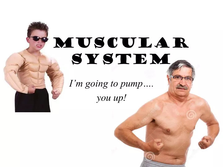
Muscular System. I’m going to pump…. you up!. Origins. Latin musculus "a muscle," literally "little mouse," The shape and movement of some muscles (notably biceps) were thought to resemble mice. Myo- is the Greek prefix. sarco- "flesh, fleshy, of the flesh,". Sarcasm.

E N D
Muscular System I’m going to pump…. you up!
Origins • Latin musculus "a muscle," literally "little mouse," • The shape and movement of some muscles (notably biceps) were thought to resemble mice. Myo- is the Greek prefix. • sarco- "flesh, fleshy, of the flesh,"
Sarcasm • Greek sarkasmos "a sneer, jest, taunt, mockery," literally "to strip off the flesh,“ Can you give me examples of sarcasm?
Muscular System Functions • Body movement (Locomotion) • Maintenance of posture • Production of body heat (Thermogenesis) • Stabilizing Joints • Respiration • Diaphragm and intercostal contractions • Communication (Verbal and Facial) • Constriction of organs and vessels • Peristalsis of intestinal tract/blood vessels; pupils • Heart beat
Properties of Muscle • Excitability: capacity of muscle to respond to a stimulus • Contractility: ability of a muscle to shorten and generate pulling force • Extensibility: muscle can be stretched back to its original length • Elasticity: ability of muscle to recoil to original resting length after stretched
Types of Muscle • Skeletal • Attached to bones (40% of body weight) • Voluntary • Smooth • In the walls of hollow organs, blood vessels, eye, glands, uterus, skin • Some functions: propel urine, mix food in digestive tract, dilating/constricting pupils, regulating blood flow, • Involuntary • Cardiac • Heart: major source of movement of blood • Involuntary
Connective Tissue Sheaths • Epimysium. Dense regular connective tissue surrounding entire muscle • Perimysium. Collagen and elastic fibers surrounding a group of muscle fibers called a fascicle • Endomysium. Loose connective tissue that surrounds individual muscle fibers
Collagen fibers of all 3 layers come together at each end of muscle to form a tendon or aponeurosis. Structure of a Skeletal Muscle
Nerve and Blood Vessel Supply • Motor neurons • stimulate muscle fibers to contract • Neuron axons branch so that each muscle fiber (muscle cell) is innervated • Form a neuromuscular junction (myoneural junction) • Capillary beds surround muscle fibers • Muscles require large amounts of energy • Extensive vascular network delivers necessary oxygen and nutrients and carries away waste.
Skeletal Muscle • Long cylindrical cells • Many nuclei per cell • Striated • Voluntary • Rapid contractions
Basic Features of a Skeletal Muscle • Muscle attachments • Most skeletal muscles run from one bone to another • One bone will move – other bone remains fixed • Origin – less movable attachment • Insertion – more movable attachment
Skeletal Muscle Structure • Composed of muscle cells (fibers), connective tissue, blood vessels, nerves • Fibers are long, cylindrical, and multinucleated • Tend to be smaller diameter in small muscles and larger in large muscles. 1 mm- 4 cm in length • Develop from myoblasts; numbers remain constant • Striated appearance • Nuclei are peripherally located
Muscle Fiber Anatomy • Myofibrils -cylindrical structures within muscle fiber • Are bundles of protein filaments (myofilaments) • Two types of myofilaments • Actin filaments (thin filaments) • Myosin filaments (thick filaments) • When myofibril shortens, muscle shortens (contracts)
Actin (Thin) Myofilaments • Thin Filament: composed of 3 major proteins • F (fibrous) actin • Tropomyosin • Troponin • Two strands of fibrous (F) actin form a double helix • Composed of G actin monomers each of which has a myosin-binding site (see yellow dot) • Tropomyosin: an elongated protein winds along the groove of the F actin double helix. • Troponin - protein which is released when there is muscle damage. In a myocardial infarction (heart attack) they look for troponin in the blood to help diagnose a heart attack.
Z line Z line
Sliding Filament Model of Contraction • Thin filaments slide past the thick ones so that the actin and myosin filaments overlap to a greater degree • In the relaxed state, thin and thick filaments overlap only slightly • Upon stimulation, myosin heads bind to actin and sliding begins
Neuromuscular Junction • Region where the motor neuron stimulates the muscle fiber • The neuromuscular junction is formed by : 1. End of motor neuron axon (axon terminal) • Terminals have small membranous sacs (synaptic vesicles) that contain the neurotransmitter acetylcholine(ACh) 2. The motor end plate of a muscle • A specific part of the sarcolemma that contains ACh receptors • Though exceedingly close, axonal ends and muscle fibers are always separated by a space called the synaptic cleft