Download
1 / 130
1.35k likes | 1.77k Views
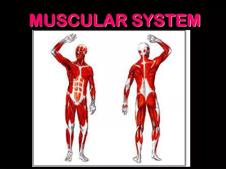
MUSCULAR SYSTEM. Muscles make up 50-60% of body wt. More than 650 muscles in the body. Each muscle is made of thousands of muscle fibers ~ the size of a fiber optic filament. It takes 17 muscles to smile & 42 to frown. The hardest working muscle is the heart.

E N D
Muscles make up 50-60% of body wt. • More than 650 muscles in the body. • Each muscle is made of thousands of muscle fibers ~ the size of a fiber optic filament. • It takes 17 muscles to smile & 42 to frown. • The hardest working muscle is the heart.
The largest muscle is the Gluteus Maximus. • The longest muscle is the Sartorius. • The strongest muscle is the Masseter
The prefix myo- & mys- means muscle & the prefix sarco- means flesh so if you hear these prefixes you’ll know we’re talking about muscles.
Muscle Functions 1. Create skeletal movement by contracting & relaxing. 2. control of organ & vessel size 3. maintain posture & position 4. support soft tissue 5. guard entrances and exits 6. maintain body temperature (85%) 7. Only body tissue that can shorten (contract)
3 Kinds of Muscle Tissue • Skeletal • Striated • Voluntary • Stacked in Sheets AKA: Somatic Tissue -Multinucleated -Found attached to skeleton
C. 3 Kinds of Muscle Tissue • Cardiacaka: heart muscle #Branched cells w/Single nuclei per cell. #Thick striations called Intercalated discsthat #Involuntary #Cells are fused so when one cell contracts, they all contract, creating the heartbeat.
3 Kinds of Muscle Tissue • Smooth aka: Visceral • Spindle shaped • Nonstriated • Involuntary -Found around hollow organs such as arteries, esophagus, stomach
D. Muscle Characteristics • Contractility Ability to shorten and exert tension or force • Excitability Ability to respond to stimuli • Extensibility Ability to contract after being stretched • Elasticity Ability to regain initial length after contraction
I. Overview E. Each muscle is an organ comprised of: • Muscle tissue (smooth, cardiac, or skeletal) • Connective tissues • Nervous tissue • Blood
II. Anatomy of Skeletal Muscle • Connective Tissue • Superficial Fascia: Fibrous connective tissue surrounding & separating each muscle
A.Connective Tissue • Deep • Epimysium – a tough outer coat of connective tissue surrounding the entire muscle. b. Perimysium - several sheathed muslce fibers wraped in a coarse fibrous membrane.
A.Connective Tissue • Deep c. Fascicles – a bundle of perimysium muscle fibers. d. Endomysium- a delicate connective sheath around a single muscle fiber
A.Connective Tissue 3. Tendons – cord like dense fibrous connective tissue. • Formed from the union of all three deep fascia • Connect muscle to muscle or muscle to bone
A.Connective Tissue 4. Aponeurosis – flat sheet of connective tissue that indirectly attaches muscles to bones, cartilage or other muscles.
B. Muscle Fibers • Each muscle fiber • is a single, long, cylindrical muscle cell. • Sarcolemma is the plasma membrane of a muscle cell. • Sarcoplasm is the cytoplasm of a muscle cell. • Many mitochondria • Nuclei • Sarcoplasmic reticulum
B. Muscle Fibers 1. Each muscle fiber • is wrapped in endomysium
1. Each muscle fiber c. is a bundle of myofibrils which is made of a budle of myofilaments
B. Muscle Fibers 2.Fascicles: • a bundle of muscle fibers • wrapped in perimysium
B. Muscle Fibers 3. Myofibrils • made of thin and thick filaments
B. Muscle Fibers 3. Myofibrils • Thick filaments made up of the protein myosin. c. Thin filaments are made up of the protein actin.
Thin filaments Tropomyosin and troponin are regulatory proteins Actin and myosin are contractile proteins.
B. Muscle Fibers 3. Myofibril d. Together, the thick and thin filaments make up the striations
B. Muscle Fibers 4. Sarcomeres- chains of tiny contractile myofibrils • Contractile unit of a muscle • Consists of overlapping thick and thin filaments Sarcomere
B. Muscle Fibers 4. Sarcomere c. Muscle contraction results from thick and thin filaments sliding past one another.
C. Neuromuscular Junction 1. Where the neuron and muscle fiber meet • The neuron and muscle fibers it controls make up a motor unit (2-2000 fibers/unit)
C. Neuromuscular Junction 3.When stimulated, all of the muscle fibers of a motor unit contract all at once.
C. Neuromuscular Junction 4. Anatomy Axon terminal – nerve end • Produces a neurotransmitter - acetycholine (Ach)
C. Neuromuscular Junction 4. Anatomy Motor end plate – site on muscle with Motor end plate acetycholine receptors Synaptic cleft - space between the nerve & motor end plate
III. Skeletal Muscle Contraction • nerve impulse • ACh released • Ach binds to receptor on muscle • Enzyme (Acetylcholine esterase removes ACh A. Initiation events
III. Skeletal Muscle Contraction • ACh causes to Na+ to diffuse into cell • If threshold is reached, action potential occurs • - impulse travels along membrane resulting in contraction B. Action Potential
III. Skeletal Muscle Contraction C. Sliding Filament Theory • Action potential causes Ca++ release from S.R • Ca++ binds to thin filament • Thin filament rotates exposing binding site for myosin • Myosin binds actin • uses ATP to "rachet" once • releases, "and binds to next actin
Calcium is the "switch" that turns muscle "on and off" (contracting and relaxing).
III. Skeletal Muscle Contraction D. How Neurotoxins Work • cobra toxin and curare • block Ach receptors • cause flaccid paralysis, potentially fatal respiratory arrest • nerve gas and insecticides • inhibit AchE • cause potentially fatal paralytic convulsions
How a Nerve Gas Works Normal Nerve Gas
Effect of Atropine on the Transmission of Acetylcholine in the presence of a nerve agent
III. Skeletal Muscle Contraction D. How Neurotoxins Work • Botulism toxin and curare • block Ach release • cause flaccid paralysis, potentially fatal respiratory arrest • Tetanus toxin • cause excessive Ach release from motor neurons • causes potentially fatal paralytic convulsions (“lock jaw”)
III. Skeletal Muscle Contraction E. Rigor Mortis • Ca++ pumps run out of ATP • Ca++ cannot be removed • continuous contraction • eventually tissues break down
A. Aerobic Respiration IV. Energy Metabolism in Sk.Ms. • Most efficient use of glucose Sources of glucose include blood glucose and stored glycogen • 36ATP/glucose • requires oxygen • occurs in mitochondria • Muscle cells have more mitochondria than any other cell • Require a steady supply of O2
B. Creatine-phosphagen system • During rest, muscles store energy as creatine phosphokinase (CPK or CK) • During intense exercise, ATP is depleted first, then CK is used to convert ADP back to ATP