Download
1 / 29
290 likes | 408 Views
Method of Study for This Section (Applied Anatomy of the Musculoskeletal System). Read assigned readings of text

E N D
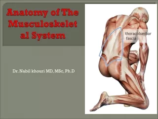
Method of Study for This Section(Applied Anatomy of the Musculoskeletal System) • Read assigned readings of text • Use the Dynamic Human CD-ROM and models and illustrations of the musculoskeletal system to help review structure of bones, joints, and muscles as necessary. This is the purpose of lab. • Complete labs conscientiously and study models of articulated skeleton, shoulder, spine, and knee • Lecture will only provide a selective review of structure, muscles & movements, and movement-related issues • For exams, review lecture notes and understanding questions in both lecture and labs
Objectives of Upper Ext Unit • Explain how anatomical structure affects movement capabilities on upper extremity articulations. • Identify factors influencing the relative mobility and stability of upper extremity movements • Identify muscles that are active during specific upper extremity movements • Describe the biomechanical contributions, specific structures, and movement-related causes of common injuries of the upper extremity.
Upper Extremity – Chapter 7 Shoulder Girdle Structure:
Normal Rom of Shoulder girdle • Sternoclavicular joint • Protraction 15 deg, retraction 15 deg • Elevation 45 deg, depression 15 deg • Acromioclavicular joint • All directions 20-30 deg
Muscles and Movement of Shoulder Girdle • Illustrations on next three slides • Trapezius (large, superficial medial and sup to scapulae) • Upper portion - elevation, upward rotation • Middle portion - adduction, or retraction • Lower portion - depression, upward rotation • Rhomboids - • elevation, downward rotation, adduction, or retraction • Serratus anterior (underneath scapulae) • abduction, upward rotation • Pectoralis minor (underneath pectoralis major) • downward rotation, abduction, or protraction • Levator Scapulae (underneath upper trapezius) • elevation, downward rotation
Normal ROM of arm movements • Flexion – (arm at side is 0 deg) 180 deg, hyperextension 60 deg • Transverse flex (arm in front of chest is 0 deg) – 40 deg, transverse ext 90 deg • Rotation(arm abducted, elbow bent to 90 deg, arm at right angles to trunk is 0 deg) • Internal 90 deg, external 90 deg • Abduction 180 deg
Shoulder Joint Stabilizers • Stabilizers and rotators - Rotator cuff muscles – • Teres minor - external rotation • Infraspinatus - external rotation • Supraspinatus - abduction • Subscapularis - internal rotation
Shoulder Joint Primary Movers • Anterior movers – Anterior deltoid, pectoralis major • Superior movers - middle deltoid • Posterior movers - posterior deltoid • Inferior movers - latissimus dorsi, teres major, lower pectoralis m. • Force vectors of muscles (see next slide)
Movements of Shoulder Complex • Every movement of upper extremity involves either stabilizing or accommodating action of the shoulder girdle. • If carrying something in arms, scapular elevators are involved • Arm elevation – scapular protraction and/or upward rotation (first 30°,1/5th is scapular movement; then 1/3rd scapular movement after that)
Shoulder Joint Impingement Syndrome • What is it? Pain from shoulder area resulting from impingement of structures between humeral head, acromion, and coracromial arch. Three stages: • Stage I - edema and hemorrhage of subacromial structures • Stage II - tendon fibrosis and bursal thickening • Stage III - rotator cuff tears, biceps tendon ruptures, and bone spurs II: III: I:
Causes of Sh Jt Impingement • Primary impingement: • Repeated movements requiring elevated and/or medially rotated humerus, compounded by weak rotator cuff muscles, causing: impingement of long head of biceps, supraspinatus • Secondary Impingement: • Decreased volume of subacromial space due to glenohumeral joint instability, and perhaps joint capsular tightness • Structural abnormalities: • hooked or curved acromion, calcium deposits, bone spurs, thickened bursa, thickened ligaments
Shoulder Jt Impingement (3) • Treatment: • Related to the cause - may involve surgery, rotator cuff strengthening, and flexibility exercises. • Later, avoid humeral elevation and rotation movements. • Website for Shoulder Joint Impingement Syndrome (click on “view eorthopod”, then “shoulder”, then “impingement syndrome”)
Elbow and Wrist Joint Muscles • True Flexor - Brachialis • Flexor-Supinator - Biceps brachii • Extensor - Triceps brachii • Wrist flexors (medial epicondyle of humerus) • Flexor carpi ulnaris and flexor carpi radialis • Wrist extensors (lateral epicondyle of humerus) • Extensor carpi ulnaris & extensor carpi radialis • Force vectors of muscles on next slide KIN 330 Biomechanics
Muscles and Movements of Radioulnar Joint • Elbow Flexion - • Forearm Supination - Biceps Brachii • Forearm Pronation - Pronator Teres • Elbow Extension - • Forearm Supination - Supinator • Forearm Pronation -Pronator Quadratus • Muscle force vectors on next slide • Epicondylitis • The most common cumulative trauma disorder (CTD), repetitive stress injury (RSI), repetitive motion disorder (RMD), or overuse syndrome (OS) is epicondylitis • Epicondylitis website (click on “view eorthopod”, then “elbow”, then “medial epicondylitis” or “lateral epicondylitis”
Normal ROM for forearm and wrist movements • Forearm • Flexion 150 deg • Supination 80-90 deg • Pronation 80-90 deg • Wrist • Flexion 80 deg • Extension 70 deg • Radial flex 20 deg • Ulnar flex 30 deg
Carpal Tunnel Syndrome • Background Carpal tunnel includes median nerve and 9 flexor tendons ( 4 flex dig sup, 4 flex dig prof, 1 fl pol l)
Carpal Tunnel Syndrome (cont’d) • Symptoms • Pain in wrist area, or referred proximally or distally • Tingling of thumb, fingers, or palmar side of hand • Loss of control of muscles affected by median nerve blockage • Causes • Enlargement of tissues within tunnel • Decreased size of tunnel • Extraneous tissue in tunnel • Treatment • Related to cause • Website on carpal tunnel syndrome (Click on “view eorthopod”, then “hand”, then “carpal tunnel syndrome” • KIN 330 Biomechanics
Review & Homework Problems for Chapter 7 • Review problems: • Torque at shoulder with elbow flexed vs extended • Fig 7-15, 7-16 • Compressive force at shoulder jt • Fig 7-17, sample problem 1 p 197 • Elbow flexion force • Figure 7-25, sample problem 2 p 206 • Homework – Due Tuesday, March 7 • Introductory problems, p 217: # 8,9,10 • Additional problem, p 218: #10