Download
1 / 37
370 likes | 385 Views
Explore the structures and functions of the respiratory system, from the nose to alveoli. Learn about ventilation mechanics, lung volumes, diffusion processes, and more in this detailed guide.

E N D
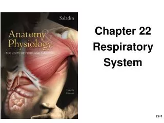
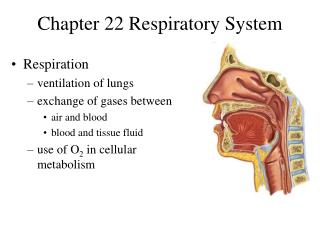
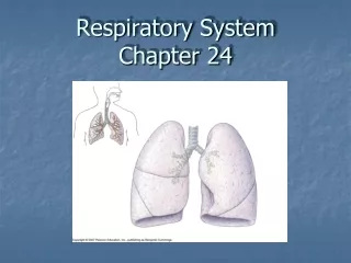
Structure and Function of the Respiratory System • Structures • Nose/nostrils • Nasal cavity • Pharynx • Larynx • Trachea • Bronchi • Bronchioles • Alveoli
Structure and Function of the Respiratory System (cont’d) • Anatomical structures of respiratory system.
Structure and Function of the Respiratory System (cont’d) • Functions • Conducts air into & out of lungs • Exchanges gases between air & blood • Humidifies air: prevents damage to membranes due to drying out • Warms air: helps maintain body temperature • Filters air • Mucus traps airborne particles • Cilia move mucus toward oral cavity to be expelled
Structure and Function of the Respiratory System (cont’d) • Alveoli • Saclike structures surrounded by capillaries in lungs • Attached to respiratory bronchioles • Site of exchange of oxygen & carbon dioxide • 300 million in lungs • Provide tremendous surface area where diffusion can take place • Respiratory membrane: 2 cell membranes that aid diffusion • Membrane of alveolar cells • Membrane of cells of capillary wall
Structure and Function of the Respiratory System (cont’d) • Respiratory membrane.
Mechanics of Ventilation • Pleural Sac • Double-layered membrane that encases each lung • Visceral (pulmonary) pleura: outer surface of lungs • Parietal pleura: inner surface of thoracic cavity & diaphragm • Pleural fluid: lubricating fluid between 2 membranes • Intrapleural pressure: pressure in pleural cavity between 2 membranes; less than atmospheric pressure
Mechanics of Ventilation (cont’d) • Pressure Changes During Ventilation • Increase in volume of intrathoracic cavity: • Increases lung volume • Decreases intrapulmonic pressure • Causes air to rush into lungs (inspiration) • Decrease in volume of intrathoracic cavity: • Decreases lung volume • Increases intrapulmonic pressure • Causes air to rush out of lungs (expiration)
Mechanics of Ventilation (cont’d) • Inspiration • Inspiratory muscles increase intrathoracic cavity volume • Diaphragm: most important inspiratory muscle • Flattens as it contracts • Puts in motion pressure changes that cause inspiration • Contraction moves abdominal contents forward & downward • Muscles that elevate ribs: external intercostals, scalenes, sternocleidomastoid, pectoralis minor
Mechanics of Ventilation (cont’d) • Expiration • No muscular effort needed at rest • Passive recoil of diaphragm & other muscles decreases intrathoracic cavity volume • During exercise or voluntary forced expiration, accessory muscles of expiration contract, pulling ribs downward: • Internal intercostals • Rectus abdominis • Internal oblique muscles of abdominal wall
Mechanics of Ventilation (cont’d) • Inspiration and expiration.
Mechanics of Ventilation (cont’d) • Muscles involved in inspiration & expiration.
Mechanics of Ventilation (cont’d) • Airflow Resistance • Airflow = P1 − P2/Resistance • Where P1 − P2 is pressure difference between 2 areas & Resistance is resistance to airflow between 2 areas • Thus, airflow can be increased by: • Amplifying pressure difference between 2 areas • Decreasing resistance to airflow • Diameter of airway is biggest factor affecting airflow at rest • In exercise, bronchodilation decreases resistance to airflow
Mechanics of Ventilation (cont’d) • Pulmonary Ventilation • Amount of air moved in & out of lungs in given time period • Tidal volume: amount of air moved per breath • Volume of air moved per minute can be calculated as: • VE = VT × f • Where VE = volume of air expired per minute; VT = tidal volume; f = breathing frequency per minute • Greater in trained athletes • Pulmonary ventilation = anatomical dead space + alveolar ventilation
Mechanics of Ventilation (cont’d) • Lung Capacities and Volumes • Determined using spirometry equipment • Reserve of tidal volume at rest allows increase in tidal volume during maximal exercise • Residual volume: air left in lungs after max. exhalation • Frequency and Depth of Breathing • Increase in depth of breathing occurs first after onset of exercise • If increase in depth not sufficient, rate of breathing will increase
Mechanics of Ventilation (cont’d) • Lung volumes and capacities.
Diffusion at the Lungs • Factors Promoting Diffusion • Large surface area of alveoli • Thinness of respiratory membrane (2 cells thick) • Pressure differences of oxygen & carbon dioxide between air in alveoli & blood • Partial pressure: portion of pressure due to a particular gas in a mixture of gases • Dalton’s law: total pressure of gas mixture = sum of partial pressures of each gas • Henry’s law: amount of gas dissolved in any fluid depends on temperature, partial pressure of gas, & solubility of gas
Diffusion at the Lungs (cont’d) • Oxygen Diffusion • Partial pressure of oxygen (PO2) must be > in alveoli than in blood & > in blood than in tissue • PO2 at sea level = 159.1 mm Hg • PO2 in alveoli = 105 mm Hg • PO2 in arterial blood entering lungs = 40 mm Hg • PO2 in blood leaving lungs = 100 mm Hg • PO2 in tissues = 40 mm Hg • Thus, differences between PO2 in alveoli & blood (65 mm Hg) and between blood & tissue (60 mm Hg) provide driving force for diffusion of oxygen
Diffusion at the Lungs (cont’d) • Capillary gas exchange at lungs & tissue.
Diffusion at the Lungs (cont’d) • Carbon Dioxide Diffusion • Partial pressure of carbon dioxide (PCO2) must be > in blood than in alveoli & > in tissue than in blood • PCO2 in atmospheric air = 0.2 mm Hg • PCO2 in alveoli = 40 mm Hg • PCO2 in arterial blood entering lungs = 46 mm Hg • PCO2 in blood leaving lungs = 40 mm Hg • PCO2 in tissues = 46 mm Hg • Thus, differences between PCO2 in alveoli & blood (6 mm Hg) and between blood & tissue (6 mm Hg) provide driving force for diffusion of carbon dioxide
Diffusion at the Lungs (cont’d) • Lung Blood Flow • Determines velocity at which blood passes through pulmonary capillaries • Increased blood flow during exercise results in increased gas diffusion • Blood pressure in pulmonary circulation is low compared with systemic • Equilibration of oxygen between alveoli air & lung capillary blood takes 0.25 seconds • As blood flow increases with exercise, less time is available for this equilibration • However, increased capillary blood volume slows blood flow
Blood Gas Transport • Oxygen Transport • Only 9 to 15 mL of oxygen can be dissolved in plasma, which is insufficient to meet needs of body • RBCs containing hemoglobin transport 98% of oxygen • Oxyhemoglobin: oxygen bound to hemoglobin • Deoxyhemoglobin: hemoglobin not bound to oxygen • Concentration of hemoglobin determines amount of oxygen that can be transported
Blood Gas Transport (cont’d) • Oxyhemoglobin disassociation curve.
Blood Gas Transport (cont’d) • Oxyhemoglobin Disassociation Curve • Temperature effect • Increase in temp. • Shifts curve to right • Decreases affinity of hemoglobin for oxygen • Decrease in temp. • Shifts curve to left • Increases affinity of hemoglobin for oxygen
Blood Gas Transport (cont’d) • Effect of temperature & acidity on hemoglobin disassociation curve.
Blood Gas Transport (cont’d) • Oxyhemoglobin Disassociation Curve (cont’d) • pH effect (Bohr effect) • Increase in acidity • Shifts curve to right • Decreases affinity of hemoglobin for oxygen • Decrease in acidity • Shifts curve to left • Increases affinity of hemoglobin for oxygen
Blood Gas Transport (cont’d) • Oxyhemoglobin Disassociation Curve (cont’d) • 2,3-Diphosphoglycerate (2,3 DPG) effect • Increase in 2,3 DPG • Shifts curve to right • Decreases affinity of hemoglobin for oxygen • Decrease in 2,3 DPG • Shifts curve to left • Increases affinity of hemoglobin for oxygen
Blood Gas Transport (cont’d) • Carbon Dioxide Transport • 3 methods • 7% to 10% is dissolved in plasma • 20% is bound to hemoglobin • 70% is transported as bicarbonate
Blood Gas Transport (cont’d) • Ability of hemoglobin to bind oxygen & carbon dioxide.
Gas Exchange at the Muscle • Occurs due to partial pressure differences between oxygen & carbon dioxide between tissue & blood • Myoglobin • Oxygen transport molecule similar to hemoglobin • Found in skeletal & cardiac muscle • Reversibly binds with oxygen • Assists in passive diffusion of oxygen from cell membrane to mitochondria • Functions as oxygen reserve at start of exercise
Control of Ventilation • Respiratory Control Center • Portion of medulla oblongata & pons • Serves as pacemaker, generating a rhythmical breathing pattern • Rate & depth of breathing can be modified by: • Higher brain centers • Chemoreceptors in medulla • Other peripheral inputs • Pulmonary ventilation is generally involuntary, but can changed voluntarily
Control of Ventilation (cont’d) • The respiratory control center in the medulla.
Control of Ventilation (cont’d) • Central Chemoreceptors • Located in medulla, separate from respiratory control center • Respond to changes within CSF, esp. in H+ concentration or pH • Peripheral Chemoreceptors • Located in carotid arteries & aortic arch • Respond to changes in blood PCO2 & H+ concentration • Other Neural Input • Stretch receptors in lungs & respiratory muscles • Proprioceptors & chemoreceptors in skeletal muscle & joints
Effects of Exercise on Pulmonary Ventilation • Three phases of changes in pulmonary ventilation.
Ventilation Is Associated With Metabolism • Ventilatory Equivalents • Amount of air ventilated needed to obtain 1 L of oxygen or expire 1 L of carbon dioxide • Ventilatory equivalent of oxygen: ratio of pulmonary ventilation (VE) to oxygen (VO2): VE/VO2 • Ventilatory equivalent of carbon dioxide: ratio of pulmonary ventilation (VE) to carbon dioxide (VCO2): VE/VCO2 • Ventilatory Threshold (VT) • Technique using ventilatory equivalents to estimate lactate threshold
Ventilation is Associated With Metabolism (cont’d) • VT and RCP.
Ventilation Is Associated With Metabolism (cont’d) • Respiratory Compensation Point (RCP) • The work intensity at which both VE/VO2 & VE/VCO2 increase • Characterized by a decrease in end-trial partial pressure of O2 • Indicates end of control of VE by PCO2 • VT & RCP can be used to create 3 training zones of exercise intensity, based on heart rate: • Light-intensity: <VT • Moderate-intensity: between VT & RCP • High-intensity: >RCP