Download
1 / 28
280 likes | 318 Views
Discover the impressive features of cartilage, a specialized connective tissue in the body that is rigid, elastic, and resilient. Learn about its composition, growth processes, types, and locations within the adult human body. Unveil the roles of chondrocytes, extracellular matrix, and different types of cartilage (hyaline, elastic, fibrous) in maintaining health and structure. Explore the vital functions and unique properties that make cartilage a fundamental tissue in the body's framework.

E N D
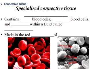
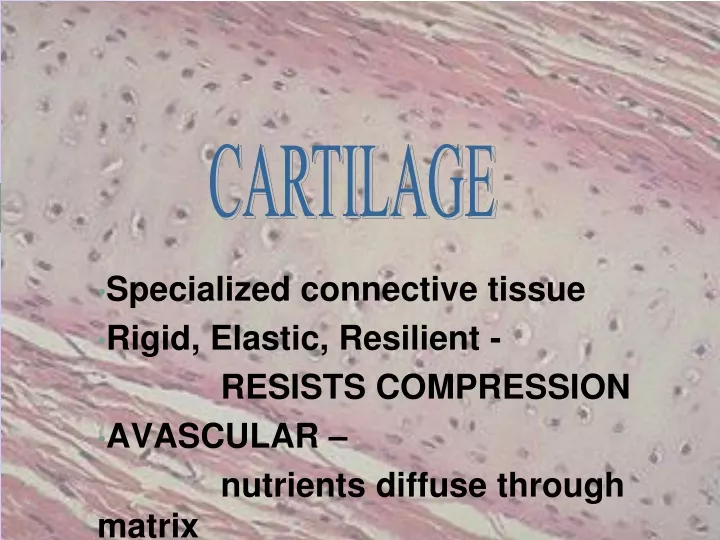
CARTILAGE Specialized connective tissue Rigid, Elastic, Resilient - RESISTS COMPRESSION AVASCULAR – nutrients diffuse through matrix
PERICHONDRIUM • Dense irregularly arranged connective tissue (type I collagen) • Ensheaths the cartilage • Houses the blood vessels that nourish chondrocytes
CHONDROBLAST • Progenitor of chondrocytes • Lines border between perichondrium and matrix • Secretes type II collagen and other ECM components • Chondroblasts build
CHONDROCYTE • Mature cartilage cell • Reside in a space called the lacuna • Clear areas = Golgi and lipid droplets
Chondrocytes completely fill their lacunae • RER and euchromatic nuclei • Synthetically active, secrete matrix N RER Cartilage matrix
MATRIX • Provides the rigidity, elasticity, & resilience • FIBERS • Collagenous and elastic • GROUND SUBSTANCE • Glycosaminoglycans (chondroitin sulfates, keratin sulfate, hyaluronic acid) • Proteoglycans: GAGs + core protein • Water • Basophilic • Territorial matrix - high [ ] of sulfated proteoglycans
CARTILAGE GROWTH • Appositional • Increasing in WIDTH; chondroblasts deposit matrix on surface of pre-existing cartilage • Interstitial • Increasing in LENGTH; chondrocytes divide and secrete matrix from w/in lacunae
Cartilage • Embryo • More prevalent than in adult • Skeleton initially mostly cartilage • Bone replaces cartilage in fetal and childhood periods
Location of cartilage in adults • External ear • Nose • “Articular” – covering the ends of most bones and movable joints • “Costal” – connecting ribs to sternum • Larynx - voice box
Epiglottis – flap keeping food out of lungs • Cartilaginous rings holding open the air tubes of the respiratory system (trachea and bronchi) • Intervertebral discs • Pubic symphysis • Articular discs such as meniscus in knee joint
Remember the four basic types of tissue… • Epithelium • Connective tissue • Connective tissue proper • Cartilage • Bone • Blood • Muscle tissue • Nervous tissue
Cartilage is connective tissue • Cells called chondrocytes • Abundant extracellular matrix • Fibers: collagen & elastin • Jellylike ground substance of complex sugar molecules • 60-80% water (responsible for the resilience) • No nerves or vessels (hyaline cartilage)
TYPES OF CARTILAGE • HYALINE • ELASTIC • FIBROUS
Types of cartilage: 3 • Hyaline cartilage: flexible and resilient • Chondrocytes appear spherical • Lacuna – cavity in matrix holding chondrocyte • Collagen the only fiber • Elastic cartilage: highly bendable • Matrix with elastic as well as collagen fibers • Epiglottis, larynx and outer ear • Fibrocartilage: resists compression and tension • Rows of thick collagen fibers alternating with rows of chondrocytes (in matrix) • Knee menisci and annunulus fibrosis of intervertebral discs
HYALINE CARTILAGE • FUNCTION • Support tissue and organs • Model for bone development • MATRIX • Type II collagen (thin fibrils) • Chondroitin sulfate, keratin sulfate, hyaluronic acid • Water • LOCATION • Tracheal rings, nasal septum, larynx, articular surfaces of joints
ELASTIC CARTILAGE • FUNCTION • Support with flexibility • MATRIX • Normal components of hyaline matrix plus ELASTIC fibers • LOCATION • External ear, external auditory canal, epiglottis • STAINS • Elastic fibers stain BLACK with Weigert stain perichondrium
FIBROCARTILAGE • FUNCTION • Support with great tensile strength • MATRIX • Type I collagen - Oriented parallel to stress plane • LOCATION • Intervertebral disks, pubic symphysis
FIBROCARTILAGE • Chondrocytes align between collagen fibers • Collagen fibers lie parallel to lines of stress
Before we look at collagen pic… • Hyaline cartilage: flexible and resilient • Chondrocytes appear spherical • Lacuna – cavity in matrix holding chondrocyte • Collagen the only fiber • Elastic cartilage: highly bendable • Matrix with elastic as well as collagen fibers • Epiglottis and larynx • Fibrocartilage: resists compression and tension • Rows of thick collagen fibers alternating with rows of chondrocytes (in matrix) • Knee menisci and annulus fibrosis of intervertebral discs
Triple helix of collagen molecules form fibril Fibrils aggregate into collagen fibers
Growth of cartilage • Appositional • “Growth from outside” • Chrondroblasts in perichondrium (external covering of cartilage) secrete matrix • Interstitial • “Growth from within” • Chondrocytes within divide and secrete new matrix • Cartilage stops growing in late teens (chrondrocytes stop dividing) • Regenerates poorly in adults
Uploaded By.... M.Farrukh Fayyaz