Download
1 / 28
280 likes | 291 Views
Dive into the levels of protein structure, amino acids, and bonding types in this informative guide. Understand primary, secondary, tertiary, and quaternary structures, as well as the functions of proteins.

E N D
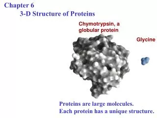
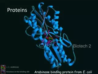
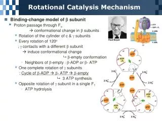
Section 2 – Protein structure, binding and conformational change • Proteins • Varied • Specific • Conformationally changed • Stability
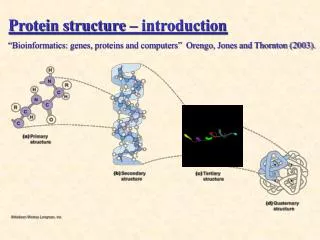
Proteins Basically they are composed of amino acids joined together but there are 4 levels to their structure: • Primary • Secondary • Tertiary • Quaternary
Basic amino acid structure carbon Amino group Carboxylic acid group Residue or Side chain
Essential amino acids Histidine, Isoleucine, Leucine, Lysine, Methionine, Phenylalanine, Threonine, Tryptophan, Valine Non-essential amino acids Alanine, Arginine, Asparagine, Aspartic acid, Cysteine, Glutamic acid, Glutamine, Glycine, Proline, Serine, Selenocysteine, Tyrosine
Proteins • 21 amino acids • Based on functional group (R group) • Properties will be in relation to the functional groups • Functional groups give 5 main amino acid groupings • Positively charged, basic • Negatively charged, acidic • Polar • Hydrophobic, non-polar • Other (special cases)(uncharged polar)
Positively charged Arginine (+NH2)
Negatively charged Aspartic acid (loses H+ on OH)
Polar (uncharged) Serine
Hydrophobic Tyrosine Phenylalanine
Other (special cases) Glycine
Bonding between amino acids in the polypeptide chain Carboxylic acid end/amide end of adjacent aa producing water Covalent peptide bond is a strong chemical bond Bond formed due to dehydration synthesis
Primary Structure Depends on the sequence of amino acids in the polypeptide chain. This is predetermined by the DNA of an organism. Only specific sequences will bring about correct function. Many amino acids joined together by peptide bonds give a polypeptide chain.
Secondary Structure Is related to how the polypeptide chain is folded. It is mainly concerned with hydrogen bonds between C=O and N-H groups. There are 2 common types of folding (with turns): -helix -sheet
H bonds between N-H groups and C=O groups. Producing helices
amino acid chains can either be parallel or antiparallel but are arranged in sheets
Tertiary Structure Relates to the folding of the alpha helices and beta sheets. This is maintained by hydrophobic interactions, H bonds, covalent disulphide bonds, ionic bonds and van der Waals interactions between side chains.
Quaternary Structure This is where 2 or more polypeptide subunits are joined together (more H bonds, ionic bonds and disulphide bridges). This also can incorporate a prosthetic non protein group such as in Haemoglobin Haemoglobin 4 polypeptide subunits and 4 haem groups
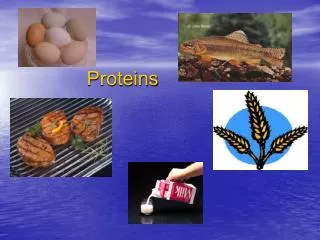
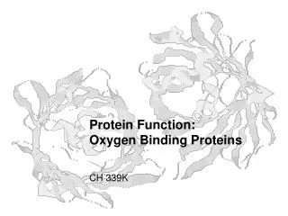
R group interactions can be influenced by temperature and pH Functions of Protein • Catalytic (enzymes) - speed up chemical reactions. • Structural - cell membrane, tissues, etc. • Messenger (hormones) - chemical messengers within cells and between cells • Carriers - proteins transporting chemicals into and out of cells.
Hydrophobic interactions Occur between non-polar R groups along the length of the polypeptide. Folding of these regions occurs so that they form a central hydrophobic core, separating non-polar hydrophobic R groups from aqueous solution while the polar hydrophilic R groups are expressed on the outside of the structure, free to interact in aqueous solution. Hydrophobic sections of proteins are classically found embedded in the phospholipid bilayer of a cell, while the hydrophilic polar parts are free to interact with the extracellular and intracellular solutions.
Hydrophobic protein domains Adenylate Cyclase is part of G protein cascade
Ionic bonds Charge dependent attraction occurring between oppositely charged polar R groups, e.g. between the amino acids arginine and aspartic acid. pH affects ionic bonding and results in denaturation of the protein at extremes of pH as the H+ and OH– ions in solution interact with the charge across the ionic bond.
Hydrogen bonds Hydrogen bonding is a weak polar interaction that occurs when an electropositive hydrogen atom is shared between two electronegative atoms. Hydrogen bonding is charge dependent. pH affects hydrogen bonding and results in denaturation of the protein at extremes of pH as the H+ and OH– ions in solution interact with the charge across the hydrogen bond.
Van der Waals interactions Weak intermolecular force between adjacent atoms. Geckos and Van der Waals forces.
Disulfide bridges Covalent bonds that form between adjacent cysteine amino acids. These can occur within a single polypeptide (tertiary structure) or between adjacent polypeptides (subunits, quaternary structure).