Download
1 / 33
590 likes | 1.73k Views
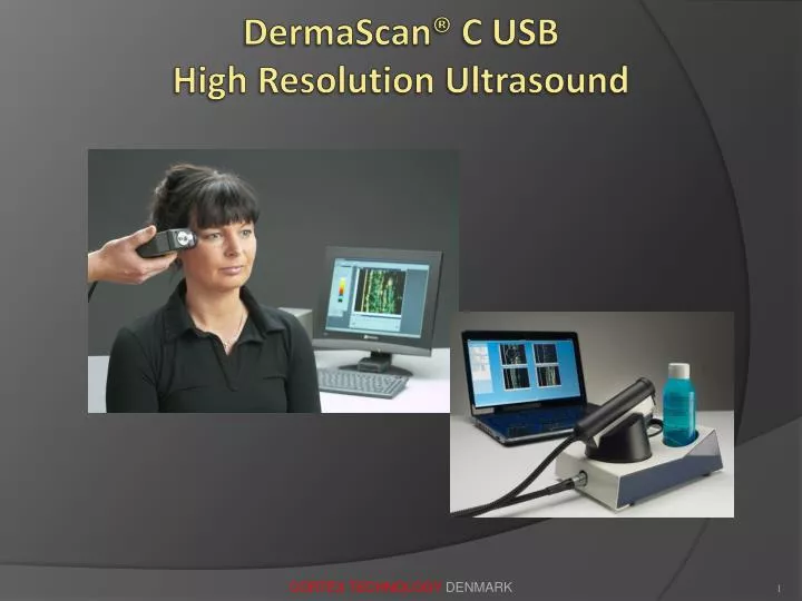
DermaScan ® C USB High Resolution Ultrasound. DermaScan® C High Resolution Ultrasound. Ultrasound Measurements - Why?. To obtain objective data To visualize invisible changes and conditions To learn more - faster To help improve treatment and life quality for patients

E N D
DermaScan® C USBHigh Resolution Ultrasound CORTEX TECHNOLOGY DENMARK
DermaScan® CHigh Resolution Ultrasound CORTEX TECHNOLOGY DENMARK
UltrasoundMeasurements - Why? • To obtain objective data • To visualize invisible changes and conditions • To learn more - faster • To help improve treatment and life quality for patients • To improve quality of products • To lower cost of product development CORTEX TECHNOLOGY DENMARK
Ultrasound Measurements - When? • Prior to therapy (e.g. tumors, port wine stains) • Follow-up (e.g. wound healing, tumors, port wine stains, aging) • To document product claims (cosmetics, pharma.) • To obtain accurate and reproducible data for basic research CORTEX TECHNOLOGY DENMARK
UltrasoundMeasurements - Who? • In dermatology & plastic surgery (clinically, research) • Pharmaceutical industry • Cosmetic and aesthetic industry • Others - neuro medicine, wound healing, paediatrics, occupational medicine etc. CORTEX TECHNOLOGY DENMARK
UltrasoundApplicationsinclude … • Scientific skin research • Irritancy/allergy testing • Wound healing • Collagen (photo damage, skin aging, skin rejuvenation) • Cellulite (efficiency of treatment) • Scleroderma, Psoriasis • Tumors (basal cell, squamouscell, malignant melanoma …) • Vascular lesions (port wine stains, hemangioma) • Claims substantiation (cosmetics and pharma.) CORTEX TECHNOLOGY DENMARK
Definition of Ultrasound • Frequency 20.000 Hz (20 kHz) Ultrasound Echograpy Sonography CORTEX TECHNOLOGY DENMARK
Conventional Ultrasound • Conventional ultrasound: fmax < 10 MHz + High penetration + Wide application range + Wide price range - Low resolution CORTEX TECHNOLOGY DENMARK
High Resolution Ultrasoundfor Skin Measurement • High resolution ultrasound: f = 20 MHz or higher + High resolution + High definition - Low penetration - Limited to skin / subcutis CORTEX TECHNOLOGY DENMARK
Send Ultrasound Pulse and listen for Echo skin boundaries CORTEX TECHNOLOGY DENMARK
The basicEchoA-Scan Skin Transmitted pulse of ultrasound Received signal (A-scan) CORTEX TECHNOLOGY DENMARK
B-Scan image = 224 lines of A-Scan Probe One image consists of 224 lines of ultrasound A-scan CORTEX TECHNOLOGY DENMARK
The B-Scan Image Epidermis Dermis Subcutis Normal face scan, young lady, near eye Probe Water Film Gel CORTEX TECHNOLOGY DENMARK
Sound beam Focal zone In-depth penetration Transducer housing Water/gel path Active element (x-tal) Skin surface BeamFocus is important for high Resolution Skin CORTEX TECHNOLOGY DENMARK
Resolution of B-Scan Image • Axial resolution (Rax) is determined by the sound velocity (v) and the system bandwidth (BW)where BW ~ 75% of FcrystalE.g. 20MHz: v ~ 1580 m/s, BW ~ 0,75 * 20 MHz => Rax ~ 60 µm50MHz: v ~ 1580 m/s, BW ~ 0,75 * 50 MHz => Rax ~20 µm • Lateral resolution is determined by the beam diameter (diameter determined by acoustic focusing) • typically 150 µm Higher ultrasound frequency higher band-width higher resolution CORTEX TECHNOLOGY DENMARK
Penetration in to the Skin • Determined by focal distance (water/gel path) • Determined by attenuation (1 dB / MHz / cm) Higher ultrasound frequency lower penetration into the skin Selection of ultrasound frequency is a trade-off between resolution and penetration CORTEX TECHNOLOGY DENMARK
DermaScan® C20 MHz <–> 50 MHz Nail foil – 20 MHz and 50 MHz Notice the higher resolution but lower penetration in the 50 MHz image CORTEX TECHNOLOGY DENMARK
DermaScan® C USB 2-D Ultrasound Imaging CORTEX TECHNOLOGY DENMARK
DermaScan® C USB2-D Ultrasound Imaging Thin and photo-aged (Steroid atrophy) Probe Malignant Melanoma Scar tissue Normal skin CORTEX TECHNOLOGY DENMARK
DermaScan® C Compact3-D Ultrasound Imgaging CORTEX TECHNOLOGY DENMARK
DermaScan® C Compact3-D Ultrasound Imaging CORTEX TECHNOLOGY DENMARK
Some examples of Images CORTEX TECHNOLOGY DENMARK
20 years 30 years 40 years 50 years DermaScan® C2-D sample Images Normal photo-aged skin Notice : The Collagen structure changes over time (colors disappear over time) CORTEX TECHNOLOGY DENMARK
DermaScan® C2-D sample Images Cellulite After AHA peeling CORTEX TECHNOLOGY DENMARK
DermaScan® C2-D sample Image Hemangioma in the face CORTEX TECHNOLOGY DENMARK
DermaScan® C2-D sample Image Scar tissue CORTEX TECHNOLOGY DENMARK
DermaScan® C2-D sample Images BCC, various body sites CORTEX TECHNOLOGY DENMARK
Measurements CORTEX TECHNOLOGY DENMARK
Ultrasound BasicsMeasurement modes • Distance (A-scan, arbitrary, B-scan edge/peak detect.) • Intensity (B-scan edge/peak detection, Region of Interest) • Area (B-scan edge/peak detection, Region of Interest) • Volume (3D-scan only) CORTEX TECHNOLOGY DENMARK
DermaScan® CDistance Measurements Insect bite CORTEX TECHNOLOGY DENMARK
DermaScan® CIntensity Measurement Insect bite CORTEX TECHNOLOGY DENMARK
DermaScan® CArea Measurement CORTEX TECHNOLOGY DENMARK
Thank You for Your Attention CORTEX TECHNOLOGY DENMARK