Download
1 / 42
420 likes | 441 Views
Uncover the fundamentals of DNA, from nucleosides to nucleotides, explore nitrogenous bases, and delve into the intricacies of DNA structure and stability. Discover the secrets encoded in the molecules of life.

E N D
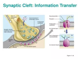
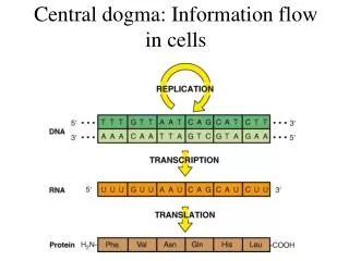
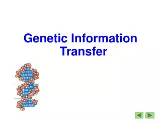
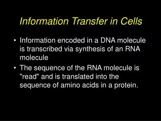
Information Transfer in Cells • Information encoded in a DNA molecule is transcribed via synthesis of an RNA molecule • The sequence of the RNA molecule is "read" and is translated into the sequence of amino acids in a protein.
Review of DNA Structure • What is a nucleoside? • What is a nucleotide? • What forces hold DNA together as a helix? • Why are there two kinds of grooves in a B DNA helix? • What are the differences between A, B and Z forms of DNA
Sugar phosphate Nitrogenous base DNA (deoxyribonucleic acid) Building blocks = deoxyribonucleotides
Nitrogenous base Phosphate 5 HoCH2 oH HoCH2 oH o o 1 4 2 3 oH Links Nucleotide units H oH oH Ribose 5 1 4 2 3 Ribose - a pentose sugar - a furanose ring - in RNA - in nucleotides for energy metabolism (ATP) 2 deoxyribose - a pentose sugar - a furanose ring - in DNA
(11.2 Pentoses of Nucleotides) • D-ribose (in RNA) • 2-deoxy-D-ribose (in DNA) • The difference - 2'-OH vs 2'-H • This difference affects secondary structure and stability
11.1 Nitrogenous Bases • Pyrimidines • Cytosine (DNA, RNA) • Uracil (RNA) • Thymine (DNA) • Purines • Adenine (DNA, RNA) • Guanine (DNA, RNA)
Properties of Pyrimidines and Purines • Keto-enol tautomerism • Strong absorbance of UV light
Guanine Guanine
Nitrogenous base 5 HoCH2 oH NH2 o 1 4 N 2 3 oH 5 H o HoCH2 N o N-glycosidic linkage 1 4 2 3 oH H Nucleoside A purine/pyrimidine + deoxyribose or ribose Cytosine 4 5 3 ‘ 6 2 1 ‘ ‘ ‘ ‘ Cytidine
11.3 Nucleosides Linkage of a base to a sugar • Base is linked via a glycosidic bond • Named by adding -idine to the root name of a pyrimidine or -osine to the root name of a purine • Sugars make nucleosides more water-soluble than free bases
11.4 Nucleotides Nucleoside phosphates • Know the nomenclature • "Nucleotide phosphate" is redundant!
NH2 N O O O 5’ o O-P-O-P-O-P-OCH2 N - - - O O O O 1’ 4’ 2’ 3’ H OH - O O -P - 5’ OCH2 Nitrogenous base O 1’ 4’ 2’ 3’ H OH Deoxyribonucleic acid DNA is a nucleotide polymer linked by a 3’ to 5’ phosphodiester bond 5’ phosphate 3’ hydroxyl
Single-stranded DNA: Has polarity Has a hydrophilic side Has a hydrophobic side
5’ 5’ 3’ 3’ Double-stranded DNA 1) Pair of DNA chains in an antiparallel arrangement 2) Sugar-P backbone outside, aromatic rings (bases) inside 3) Bases pair specifically by H-bonding A pairs with T; G pairs with C [A] = [T] and [G] = [C] [purines] = [pyrimidines]
The “canonical” base pairs • The canonical A:T and G:C base pairs have nearly identical overall dimensions • A and T share two H-bonds • G and C share three H-bonds • G:C-rich regions of DNA are more stable • Polar atoms in the sugar-phosphate backbone also form H-bonds
Why a helix? Why not a ladder? • A side view of base pairs shows they are perpendicular to the helix axis • The heterocyclic bases have flat surfaces which are hydrophobic • To exclude water from between the rings, we should bring the bases closer together • One way to model them closer together is to “twist” the ladder into a helix
Right-handed twist ~10 base pairs per turn B form DNA helix
Summary: What holds DNA together? • Sugar-phosphate backbone outside • (1) minimizes electrostatic repulsion, • (2) interacts with water • Bases inside • (3) hydrogen-bonded • (4) plus base stacking by hydrophobic interactions
Major and minor grooves • The "tops" of the bases (as we draw them) line the "floor" of the major groove • The major groove is large enough to accommodate an alpha helix from a protein • Regulatory proteins (transcription factors) can recognize the pattern of bases and H-bonding possibilities in the major groove
Comparison of A, B, Z DNA • A: right-handed, short and broad, pitch is 2.3 A, 11 bp per turn • B: right-handed, longer, thinner, pitch is ~3.4 A, ~10 bp per turn • Z: left-handed, longest, thinnest, pitch is 3.8 A, 12 bp per turn
DNA Packaging • Human DNA total length is ~2 meters • Is packaged into a nucleus that is ~ 5 microns in diameter • This represents a compression of more than 100,000 fold • It is made possible by wrapping the DNA around protein spools called nucleosomes and then packing these into helical filaments
We reviewed: Chapter 11, Sections: 11.1, 11.2, 11.3, 11.4, 11.5 and the “DNA parts” of 11.6 Chapter 12, Sections: 12.2, 12.5