Download
1 / 48
480 likes | 497 Views
Learn about the application of Metal and Hybrid TimePix Detectors in measuring and imaging low-energy ion beams at various stages of the laser mass-spectrometer. Explore the future of Metal Micro-detectors with TimePix for beam analysis.

E N D
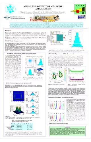
Metal and Hybrid TimePix DetectorsImaging Beams of Particles Valery Pugatch Institute for Nuclear Research National Academy of Sciences Kiev, Ukraine & M. Campbell, L. Tlustos, X.Llopart , CERN S. Pospisil, IEAP, Prague V. Storizhko, V. Eremenko, S. Homenko, A. Shelekhov, IAP NASU, Sumy A. Chaus,O.Kovalchuk, O. Okhrimenko, D. Storozhik, INR NASU, Kiev PIXEL2010. Grindelwald. September 6-10, 2010 PIXEL2010. V. Pugatch.
Content • Introduction • Hybrid (Silicon) TimePix measuring and imaging low energy ion beams: • at the focal plane of the laser mass-spectrometer • at the ion source • Metal TimePix Detector: • Metal detectors of radiation – physics and technique. • Measuring and imaging low energy ion beams at the focal plane of the laser mass-spectrometer • FUTURE - Metal Micro-detectors readout by TimePix for measuring and imaging beams • Summary and Outlook. PIXEL2010. V. Pugatch.
Institute for Nuclear Research National Academy of Sciences Kiev, Ukraine. 5 Sections: • Nuclear Physics(HEP Department including) • Atomic Energy • Solid-State Physics • Plasma Physics • Radiobiology and Radioecology • Facilities:Isochronous Cyclotron U-240,Tandem ЕSG-10К,Cyclotron U-120,Nuclear Reactor WWR-M. Isochronous cyclotron beams: • Protons up to 100 MeV • Deuterons – up to 60 MeV • Alpha – 120 MeV • Heavy ions (Ne, Ar, Xe) – 120 Z2/A MeV/nucleon PIXEL2010. V. Pugatch.
Introduction • Recent years - significant achievements in miniaturization of mass spectrometers. Many areas of science and technology need such devices providing simultaneous measurement of wide mass-spectra in real time . • This progress occurred, in particular, due to micro-detectors and readout electronics developments for High Energy Physics experiments. • Microstrip as well as micropixel detectors could be applied to create the Electronic Focal Plane (EFP) for mass spectrometers. PIXEL2010. V. Pugatch.
Introduction (continued) • EFP - Current status: SEM, MCP, Micro-Faraday cup arrays etc., • Requirements: • Large dynamic range: up to 107 – 108 • Sensitivity to a single ion • Real time data simultaneously over all chemical elements • These studies: TimePix evaluation for measuring and imaging low energy ions beams: • at the focal plane of the laser mass-spectrometer • at the ion source : Hybrid & Metal - response over 256 x 256 pixels. Future radiation hard Metal Micro-detectors+TimePix for ion and synchrotron beams PIXEL2010. V. Pugatch.
Hybrid (Silicon) TimePix • measuring and imaging low energy ion beams: • at the focal plane of the laser mass-spectrometer • at the ion source The goal of the studies: • TimePix response as a function of: • Ion energy (3 – 40 keV) • Ion mass (H, He, Al, Cu, Zr, Nb, Sn, Ta, Pb) • Ion charge (1+, 2+, 3+, 4+) • Ion beam position over 256 x 256 pixels. PIXEL2010. V. Pugatch.
Hybrid pixel detector with the n-Silicon sensor chip and the TimePix electronics chip connected viabump bonds. Hybrid (Silicon) TimePixat the focal plane of the laser mass-spectrometer L.Tlustos PIXEL2010. V. Pugatch.
Schematics of the ion path in a laser mass-spectrometer.(Institute of Applied Physics NASU, Sumy, Ukraine.) Ions: H – Pb, 1+ <= Z <= 4+, 3 – 80 keV. 1 – Laser, 2 – Target, 3 – Accelerator, 7 – energy analyzer, 9 – Magnet, 10 – TimePix, 11 – Focal plane The TimePix chip was mounted in a vacuum chamber on a moveable platform at the focal plane of a laser double-focusing mass spectrometer. PIXEL2010. V. Pugatch.
TimePix at the focal plane of the Laser Mass Spectrometer. PIXEL2010. V. Pugatch. Ужгород, 17-18 травня 2007
TimePix at the focal plane of the laser mass-spectrometer • Ions beam has been generated at the sample-target by the infrared (1064 nm) laser (15 ns, 50 Hz). • Passing through the magnetic sector ions were focused accordingly to their mass over charge ratio in a focal plane (210 mm long) of the mass-spectrometer. • For each bunch of ions detected by a pixel a triangular pulse is formed with a height proportional to a number of ions in a bunch. Whenever the new bunch of ions arrives at the pixel its counter content is increased accordingly to the number of ions in the bunch. • TimePix chip was readout by the PIXELMAN hardware/software (IEAP, Prague) via USB-connection to PC. • Real-time digital information, high speed communication and data transfer are essential features of TimePix chips for a mass-spectrometry. PIXEL2010. V. Pugatch.
TimePix at the focal plane of the Laser Mass Spectrometer. TOT mode - For each bunch of ionsdetected by a pixel a triangular pulse is formed with a height proportional to a number of ions in a bunch. Whenever the new bunch of ions arrives at the pixel its counter content is increased accordingly to the number of ions in the bunch. The effect of the non-linearity at the TOT value lower than 6.5 μs has to be taken into account for obtaining correct mass-spectrum These studies: THL = 390 – 395, Integration – ( 0.1 – 5) s PIXEL2010. V. Pugatch.
Studies performed (July 2008 – August 2010) TimePix (Hybrid and Metal) – 2 & 2 samples. Uniformity of response over : • ion energy (3 - 40 keV) • ion charge (1+ – 4+) and mass (H- Pb) • position of an ion beam at the detector surface PIXEL2010. V. Pugatch.
Hybrid (Silicon) TimePix at the focal plane of the laser mass-spectrometer X – axis – along the focal plane (mass-spectrum) Y – axis – along the image of the laser beam spot at the target Z – axis – intensity of the analyzed ions 2D mass spectrum. Position of the TimePix, Accelerating voltage, Magnetic field adjusted – to observe 12 keV 2+ ions of Pb isotopes • Two dimensional data on-line – ‘electronic photo-plate’ – for alignment, focusing, verifying stability of electric and magnetic fields etc.,) • A powerful tool in a feedback system for fine tuning of a mass-spectrometer and similar devices. PIXEL2010. V. Pugatch.
Hybrid (Silicon) TimePix at the focal plane of the laser mass-spectrometer Mass-spectra obtained by projection of the 2-D distribution onto the mass axis. Resolution is good enough to separate single mass unit different lead isotopes. PIXEL2010. V. Pugatch.
TimePix (Hybrid-1) - response over 256 x 256 pixels. Review of the results measured by two samples of the TimePix (Hybrid) Copper isotope peaks observed at three pixel regions (0.0, 2.8 and 5.6 mm along the focal plane) by increasing the magnetic field. The ratio of 63Cu/65Cu varies by the order of magnitude (Sample 1) PIXEL2010. V. Pugatch.
TimePix (Hybrid-1). Position dependence of the pixels response: 63,65Cu and Ta single-charged ions. 65Cu 63Cu Order of magnitude non-uniformity over different regions of the TimePix chip (Hybrid) – Sample 1. Ta PIXEL2010. V. Pugatch.
TimePix (Hybrid-1) response uniformity • Al-ions, 1+, E = 12.1 keV (Sample 1). • 9 areas - (85 x 85) pixels each: • TOP, MIDDLE and BOTTOM • part of the TimePix surface PIXEL2010. V. Pugatch.
Uniformity of the TimePix (Hybrid-1) response. Zr/Nb – isotopes mass-distribution for 1+ - 4+ ions. Non-uniform response (Sample 1) dependent on the ion charge and position. PIXEL2010. V. Pugatch.
Uniformity of the TimePix (Hybrid-1) response. Zr/Nb – isotopes mass-distribution for 1+ - 4+ ions. Acceleration voltage = 12.1 kV Uniformity of response is a crucial feature for a sensor imaging mass distribution. Results obtained for the TimePix (Sample1) show irregularity in response by different pixels. PIXEL2010. V. Pugatch.
Irregularities in TimePix (Hybrid-1)-response to 14C – beta-particles 14C. Depletion voltage – 0 V. Order of magnitude difference in response 14C. Depletion voltage – 100 V. Factor of 4 – difference in response. PIXEL2010. V. Pugatch.
Aperture: 3 holes, 1mm diameter separated by 4 mm Response of MEDIPIX-2 (Hybrid-1) to low energy ions. Factor of 3 difference in the response of MEDIPIX-2 (Hybrid) to He and H2 ions of the same energy. Ion source at the KINR isochronous cyclotron U-240.The response variation over the surface of the detector (Sample 1) exceeds order of magnitude.The most probable reason – varying thickness of the Si-sensor dead layer (0.1 μm - range). Energy of ions – 30 keV. PIXEL2010. V. Pugatch.
Medipix-2 (Hybrid-1) response to a sunlight - affected by the irradiation of ions at U-240 – thin layers of Al – were deposited (~ 50 nm). Preliminary conclusion: non-uniformity of the response is determined by non-uniformity of the dead-layer thickness over the Medipix-2 (hybrid) PIXEL2010. V. Pugatch.
TimePix (Hybrid-2) response uniformity TimePix (Hybrid) – Sample 2. Zr - ions,2+, E = 19 keV Perfect (+/- 1 %) uniformity of the response ! Table: http://en.wikipedia.org/wiki/Zirconium PIXEL2010. V. Pugatch.
TimePix (Hybrid-2) response uniformity TimePix (Hybrid) – Sample 2. Zr - ions,2+, E = 25.2 keV+/- 1 % uniformity of the response ! PIXEL2010. V. Pugatch.
TimePix (Hybrid-2) Response dependence on energy and beam position (Top, Middle, Bottom area of the chip) for Zr – 2+ ions. PIXEL2010. V. Pugatch.
TimePix (Hybrid-2) ‘Charge quantum’ dependence on the ion energy. Zr (2 +). PIXEL2010. V. Pugatch.
TimePix – Metal Detector Test of the TimePix readout chip as a metal detector at the focal plane of the laser mass-spectrometer Photo of the individual pixels of the TimePIx chip (55 x 55 μm2, 256 x 256 pixels) CERN, MEDIPIX Collaboration. Diameter of the area for bonding ~15 μm. A positive voltage has been applied to a mesh over the chip area – to collect Secondary electrons emitted by metal surface of the chip under the impinging low energy ions at the focal plane. PIXEL2010. V. Pugatch.
Metal Detectors. Physics principle. PIXEL2010. V. Pugatch.
Metal Detector. Technical realization. Metal Foil Detector technology allows for Building any size beam monitoring systems: HERA-B Luminosity monitoring, LHCb Radiation Monitoring system BPM for 21 MeV proton beam (tandem MPIfK) BPM for the LHCb (ST) test beam studies 19 keV Synchrotron BPM at HASYLAB 5 MeV Electron beam BPM - KINR PIXEL2010. V. Pugatch.
Metal Detectors at LHCb Radiation Monitoring System Silicon Tracker, LHCb. M.I.P. PIXEL2010. V. Pugatch.
TimePix – Metal Detectorin a focal plane of the laser mass-spectrometer. 20th May. 2009 TimePixMetal performance for low energy ions 14th Nov. 2009 The TimePix (Sample1) was used to improve focusing: the vertical size is ~ 3 times less, now! PIXEL2010. V. Pugatch.
TimePix (METAL-1)in a focal plane of the laser mass-spectrometer. 2D mass spectra of sample with Zr2+ – isotopes . Energy of ions 12,3 keV Two dimensional data on-line – ‘electronic photo-plate’ – for alignment, focusing, testing stability of electric and magnetic fields etc.,) A powerful tool in a feedback system for fine tuning of a mass-spectrometer and similar devices. X – axis – along the focal plane (mass-spectrum) Y – axis – along the image of the laser beam spot at the target Z – axis – intensity of the analyzed ions PIXEL2010. V. Pugatch.
TimePix (METAL-1)in a focal plane of the laser mass-spectrometer. Table: http://en.wikipedia.org/wiki/Zirconium Suppression of the low intensity beam (96Zr) – non-linear TOT operation at low input charge 1 % response variation of TimePix (Sample1) different pixels PIXEL2010. V. Pugatch.
Response of the TimePix (Metal-1) to Cu isotopes as a function of ion (2+) energy PIXEL2010. V. Pugatch.
Comparison of the TimePix Hybrid/Metal sensitivity to low energy ions • The number of Zr (2+) ions per 1 count of the TimePix (Hybrid-2) at 25.2 keV is ~ 6 • The number of Zr (2+) ions per 1 count of the TimePix (Metal-2) at 25.2 keV is ~ 6 000 • Geometry factor (ratio of active sensor surface) - ~ 30 • Thus, Hybrid/Metal factor of sensitivity is ~ 33. Very thin surface layer of the silicon sensor contributes. PIXEL2010. V. Pugatch.
TimePix (METAL-2)in the focal plane of the laser mass-spectrometer Mapping isotope population over the sample area by scanning it with laser or charged particle micro-beam might be useful for applications in material technology, microbiology, medical diagnostics etc. TimePix (Metal-2) Response at different positions of 2+ ion beams (Superimposed data of three measurements) PIXEL2010. V. Pugatch.
TimePix (METAL-2)in the focal plane of the laser mass-spectrometer TimePix (METAL-2) response at different positions of ion (Charge 2+ and 3 +) beams and operating voltage (0 and 70 V) PIXEL2010. V. Pugatch.
TimePix (METAL-2)in the focal plane of the laser mass-spectrometer Udet = 70 V Table: http://en.wikipedia.org/wiki/Zirconium Suppression of the low intensity beam (96Zr) – result of the non-linear TOT operation at low input charge PIXEL2010. V. Pugatch.
TimePix(METAL-2)in a focal plane of the laser mass-spectrometer Udet = 0 V Suppression of the low intensity beam (96Zr) – the result of the non-linear TOT operation at low input charge Table: http://en.wikipedia.org/wiki/Zirconium PIXEL2010. V. Pugatch.
TimePix (METAL-2)in the focal plane of the laser mass-spectrometer Image of the grid wires • Wires diameter – 50 μm PIXEL2010. V. Pugatch.
Future work • Test a prototype of the Electronic focal plane: • 5 TpX in collaboration with C. Ponchut (ESRF) • + MCP . Single ion detection sensitivity at few keV energy 27Al (1 +) –ions has been demonstrated for MCP + CMOS active pixel sensors PIXEL2010. V. Pugatch.
Future: Electronic Focal Plane . • Metal Strip & Pixel Detectors – • for Beam Profile Monitoring • at the submicron level. • Currently available – • 1024 strips metal detector, • 60 μm pitch. PIXEL2010. V. Pugatch.
Future: Metal Micro Detectors for BPM • Advantages of the MMD: • High Radiation tolerance (10-100 MGy) • Nearly transparent sensor – 1 μm thickness • Low operation voltage (20 V) • Spatial resolution (5 – 25 μm) • Well advanced production technology • Commercially available readout hardware • and software. MMD- 16 sector pixels MMD-64. PIXEL2010. V. Pugatch.
MMD could be helpful in Particle Therapy • MMD – an ideal monitoring device: • present in the beam, permanently • does not disturb a beam • measures X-Y dose distribution • provides self-control of functionality • operates reliable over high doses From : Ken Peach (UK)Summary talk. “ Physics for Health in Europe”. CERN, 2010 PIXEL2010. V. Pugatch.
SUMMARY and OUTLOOK • The TimePix (Hybrid and Metal) response to low (3 – 40 keV) energy ions has been studied as a function of ion mass, charge, energy and beam position. • The TimePix (Hybrid) chips have demonstrated sample dependence of the pixels response. The pixels response is more uniform (+/- 1 %) for the TimePix (Metal) mode operation. • The possibility of building electronic focal plane of the mass-spectrometer based on the TimePix detector operated in a metal mode has been demonstrated. • The TimePix (Metal & Hybrid) (5 chips systems) will be evaluated soon as a demonstrator of the 'Electronic Focal Plane' . • Metal micro-detectors are on the way to become super-thin (transparent) radiation hard detectors for measurements of intensity distribution of the particles beams. PIXEL2010. V. Pugatch.
Acknowledgements • We would like to thank • MEDIPIX Collaboration • Valkov & Kolessov – KINR, U-240 team. • This work was funded partially by the CNCP (UK) project “Microstrip Metal Detectors”. PIXEL2010. V. Pugatch.
Thank you for your attention ! PIXEL2010. V. Pugatch.