Download
1 / 79
820 likes | 1.07k Views
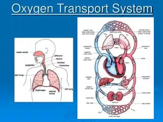
Oxygen Transport Systems. Integration of Ventilation, Cardiac, and Circulatory Functions. Cardiovascular Function. transportation of O 2 and CO 2 transportation of nutrients/waste products distribution of hormones thermoregulation maintenance of blood pressure.

E N D
Oxygen Transport Systems Integration of Ventilation, Cardiac, and Circulatory Functions
Cardiovascular Function • transportation of O2 and CO2 • transportation of nutrients/waste products • distribution of hormones • thermoregulation • maintenance of blood pressure
Cardiac Fibers Develop Graded Tension • Frank-Starling Law of the Heart • graded Ca2+ release from SR • dependent on Ca2+ influx through DHP channels
Autorhythmic cells depolarize spontaneously leaky membrane SA and AV node
Group III Central command input and output
Cardiac output affected by: • preload – end diastolic pressure (amount of myocardial stretch) • affected by venous return • afterload – resistance blood encounters as it leaves ventricles • affected by arterial BP • contractility – strength of cardiac contraction • heart rate
Mechanisms affecting HRVO2 = HR SV (a-v O2) Sinoatrial node is pacemaker for heart • spontaneously depolarizes • leakiness to Na+ • influenced by autonomic NS • training down-regulates ß-adrenergic system causing bradycardia
Cardiac Output Regulation Extrinsic control • autonomic nervous system • sympathetic NS (1 control at HR >100 bpm) • parasympathetic NS (1 control at HR <100 bpm) • stimulates ß-adrenergic receptors on myocardium • hormonal • EPI, NE
Mechanisms affecting SVVO2 = [HR SV] (a-v O2) • amount of Ca2+ influx • APs open Ca2+ channels on t-tubules • also stimulates Ca2+ release from SR • length-tension relationship • [Ca2+]-tension relationship • ß1-adrenergic modulation • activates cAMP phosphorylates L-type Ca2+, SR Ca2+ channels and pumps, troponin • Ca2+ influx and Ca2+ release from SR • training LV EDV
Intrinsic control • Frank-Starling Principle • Ca2+ influx w/ myocardial stretch • stretched fibers work at optimal length-tension curve Dotted lines indicate end-systole and end-diastole
Cardiovascular Response to Exercise Laughlin, M.H. Cardiovascular responses to exercise. Adv. Physiol. Educ. 22(1): S244-S259, 1999. [available on-line]
Cardiovascular Response to Exercise Fick principle VO2 = Q (CaO2 – CvO2) VO2 = [HR SV] (CaO2 – CvO2) VO2 = [BP TPR] (CaO2 – CvO2)
Exercise Effects on Cardiac Output • HR caused by • sympathetic innervation • parasympathetic innervation • release of catecholamines • SV, caused by • sympathetic innervation • venous return
Myocardial Mechanisms Influencing SV During Exercise • SV = EDV – ESV • Factors that influence SV • Heart size (LVV) • LV compliance during diastole • Progressive in ESV with graded exercise is from contractility • Attributed to sympathetic NS, length-tension changes • Influx of Ca2+ through L-type Ca2+ channels stimulates Ca2+ from SR release channels (Ca2+-induced Ca2+-release)
Role of Ca2+ in Cardiac Function • influx of Ca2+ through L-type Ca2+ channels stimulates Ca2+ from SR release channels (Ca2+-induced Ca2+-release) • amount of Ca2+ released from SR dependent on sarcomere length • SERCA pumps return Ca2+ to sarcoplasmic reticulum • sympathetic -adrenergic stimulation contractile force and relaxation time • affects Ca2+ sensitivity through phosphorylation • increases length of diastole to filling time
SV during graded running Zhou et al., MSSE, 2001
Effect of training and maximal exercise on VO2, Q, and a-v O2 difference
Effect of training and maximal exercise on VO2, Q, and a-v O2 difference
Arterioles and Capillaries • arterioles terminal arterioles (TA) capillaries collecting venules (CV) • arterioles regulate circulation into tissues • under sympathetic and local control • precapillary sphincters fine tune circulation within tissue • under local control • capillary density 1 determinant of O2 diffusion
Regulation of Blood Flow and Pressure Blood flow and pressure determined by: A. Vessel resistance (e.g. diameter) to blood flow B. Pressure difference between two ends A cardiac output arterioles B A B
Control of Blood Flow Blood flow to working muscle increases linearly with muscle VO2
(1-adrenergic receptor blocker) 30 s Onset of exercise
Local Control of Microcirculation • metabolic factors that cause local vasodilation • PO2 • PCO2 • H+ • adenosine • endothelial factors that cause local vasodilation • nitric oxide (NO) • released with shear stress and EPI • redistributed from Hb—greater O2 release from Hb induces NO release as well
Adenosine metabolism in myocytes and endothelial cells ATP ADP AMP adenosine Adenosine is released in response to hypoxia, ischemia, or increased metabolic work
Single layer of endothelial cells line innermost portion of arterioles that releases nitric oxide (NO) causing vasodilation
Hemoglobin • consists of four O2-binding heme (iron containing) molecules • combines reversible w/ O2 (oxy-hemoglobin) • Bohr Effect – O2 binding affected by • PO2 • PCO2 • pH • temperature • 2,3-DPG (diphosphoglycerate)
Factors affecting Oxygen Extraction Fick principle VO2 = Q (CaO2 – CvO2)
O2 extraction during graded exercise Sympathetic stimulation causes spleen to constrict releasing RBC into blood, thus increasing O2-carrying capacity
Bohr effect on oxyhemoglobin dissociation • PO2, pH and PCO2, temperature, and 2,3 DPG shift curve to left causing greater O2 release