Download
1 / 29
290 likes | 339 Views
Learn about the brain's major structures, functions, cranial nerves, meninges, CSF flow, BBB, brain subdivisions, and more. Understand the gray and white matter organization, cerebral cortex, basal nuclei, and diencephalon functions. Enhance your knowledge of brainstem, cerebellum, medulla oblongata, and cranial nerves roles. Dive deep into brain anatomy and functionality in this informative lecture.

E N D
Lecture on Brain and Cranial Nerves www.AssignmentPoint.com
Ch 15: Brain and Cranial Nerves Discuss the organization of the brain, including the major structures and their functions Describe the meninges of the spinal cord and brain, and integrate the formation and flow of CSF with this information. Describe the structures that constitute the BBB and their functions Review the cranial nerves, again giving a brief function of each.
Major Brain Subdivisions • Telencephalon (= Cerebrum) • Diencephalon (Thalamus and hypothalamus) • Mesencephalon • Metencephalon (Pons and cerebellum) • Myelencephalon (= Medulla oblongata) Brainstem
Gray & White Matter Organization In brain stem similar to spinal cord (nuclei around ventricles, tracts on outside) In cerebrum and cerebellum: white matter covered with layer of neural cortex (grey)
Cranial Meninges 2. Arachnoid - spidery, holds blood vessels 3. Pia mater - "delicate mother" 1. Dura mater - strong, "tough mother" a. falx cerebri b. falx cerebelli c. tentorum cerebelli
Longitudinal fissure Arachnoid granulations: This is where the CSF produced in the choroid plexuses of the ventricles and which has circulated into the subarachnoid space is reabsorbed.
Four Ventricles CSF filled chambers Communicating with central canal of spinal cord Lined by ependymal cells
Formation in ventricles by specialized ependymal cells of choroid plexuses(~500 mL/day; total volume ~ 150 mL) Functions transport medium, in shock absorption buoyancy(floats the brain) CSF circulation: Ventricles → central canal → subarachnoid space Reabsorption into circulation via arachnoid granulations into superior sagittal sinus. CSF: Cerebro-Spinal Fluid Fig 15.6
Blood Brain Barrier (BBB) what is it? 3 areas in brain don’t have BBB • portion of hypothalamus • pineal gland (in diencephalon) • choroid plexus
Cerebrum • Two hemispheres separated by longitudinal fissure • Gyrus (gyri) separated by sulcus (sulci) • Major lobes named after overlaying bones
Cerebral Hemispheres . . . • . . have functional regions (motor, sensory and association areas) • . . . have some functional differences (in spite of anatomical resemblance) → Lateralization of cortical functioning • . . . receive information and generate commands for opposite side of body
Cerebral Cortex and Central White Matter Gray surface (cortex) with white tracts internally Commissures – connect corresponding gyri of the two hemispheres 1) corpus callosum 2) anterior commissure Projection tracts (fibers) – connect more or less vertically Association tracts (fibers) – connect one gyrus to another in the same hemisphere
Basal (or cerebral) Nuclei Misnomer: basal ganglia Gray matter internal to the cerebral cortex, below floor of lateral ventricles. Function: modulate motor output from the cerebral cortex. Subconscious control of skeletal muscle tone and coordination of learned movement patterns. Parkinson's disease is caused by the loss of at least 80% of the dopaminergic neurons in basal nuclei and substantia nigra (resting tremor) Fig 15.11
Diencephalon Epithalamus Pineal gland - produces melatonin, sets diurnal cycles Thalamus (~12 nuclei) Hypothalamus Just superior to optic chiasma Infundibulum - connects to pituitary gland Some functions: Control of autonomic nervous system Coordination of nervous and endocrine systems Secretion of hormones - ADH and oxytocin
Mesencephalon) = Midbrain Corpora quadrigemina = 2 pairs of sensory nuclei • Superior colliculi (relay station for visual information) • Inferior colliculi (relay station for auditory information Substantia nigra - regulates motor output Cerebral peduncles - ascending and descending tracts to thalamus Nuclei of ori for CN III and IV
Metencephalon: Cerebellum Hemispheres and lobes Cortex -gray surface with folia - fine ridges and sulci - grooves between the ridges Purkinje cells , axons of which become arbor vitae (white matter) in center Regulation of posture and balance
Metencephalon: PonsMyelencephalon: Medulla oblongata • Mostly ascending and descending tracts • Nuclei of ori for many cranial nerves • Location of autonomic nuclei involved in respiratory and cardiovascular control • Relay stations for sensory and motor neurons
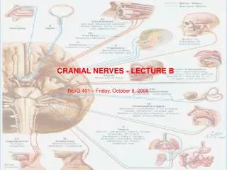
Cranial Nerves • Twelve pairs: • 2 attach to forebrain (Telen- & Diencephalon) • 10 attach to brainstem (Mes-, Met- and Myelencephalon) • Names relate to appearance or function • Classification ?
Olfactory Nerve (= CN or N I) 1º function? Origin? Destination? _____________(By way of cribiform plate of ethmoid) Only CN directly attached to Cerebrum
Optic Nerve (N II) 1º fu? ori? dest? - by way of optic foramen of sphenoid to Diencephalon (optic chiasma) and to occipital lobe
Oculomotor (N III) C: Motor O: Mesencephalon D: Somatic motor to superior, inferior, medial recti and inferior oblique; visceral motor to intrinsic eye muscles by way of superior orbital fissure
Trochlear (N IV) C: Motor O: Mesencephalon D: superior oblique by way of superior orbital fissure
Trigeminal (N V) C: Mixed three major branches 1. ophthalmic (sensory) 2. Maxillary (sensory) 3. Mandibular (mixed) O: face / nuclei of pons D: sensory nuclei in pons / muscles of mastication
C: Motor O: Pons D: Runs lateral rectus eye muscle Abducens(CN VI)
Facial (N VII) C: Mixed O: sensory from taste receptors of anterior 2/3 of tongue / motor from pons D: Sensory to sensory nuclei of pons / motor muscles of facial expression, visceral motor to tear gland.
C O ? D Vestibulocochlear (N VIII)
Glossopharyngeal (CN IX) C: mixed O: sensory from posterior 1/3 of tongue / motor from medulla oblongata D: medulla / muscles for swallowing, parotid gland
Vagus (N X) C: Mixed O: Sensation from pharyngeal area and outer ear / motor from medulla D: Sensory to medulla / visceral motor to thoracic and abdominal cavities and their organs. Major motor pathway for ANS
Accessory (N XI) and C: Motor O: Motor nuclei of medulla and spinal cord D: Swallowing, trapezius & scm muscles Hypoglossal (N XII) C: Motor O: Motor nuclei of medulla D: Tongue musculature