Download
1 / 27
1.98k likes | 6.46k Views
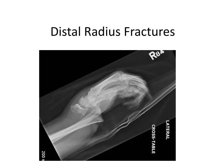
Distal Radius Fractures. Distal Radius Anatomy. Radial Styloid Lister’s Tubercle Ulnar styloid DRUJ. Radiographic Anatomy. Radial Inclination – 22 degrees Radial Ht – 11 mm Volar Tilt – 11 degrees Ulnar Varience. RADIAL Height (x) = ~ 11 – 12 MM; INCLINATION = ~ 22 0.

E N D
Distal Radius Anatomy • Radial Styloid • Lister’s Tubercle • Ulnar styloid • DRUJ
Radiographic Anatomy • Radial Inclination – 22 degrees • Radial Ht – 11 mm • Volar Tilt – 11 degrees • Ulnar Varience
RADIAL Height (x) = ~ 11 – 12 MM; INCLINATION = ~ 220 VOLAR TILT = ~ 110
What is Ulnar Variance? Ulnar Negative Ulnar Positive
Radiographic Anatomy • MRN 34611947 • Draw lines using PACS
Fracture Subtypes of the Distal Radius • Colles • Smiths • Bartons – Volar and Dorsal • Chauffeur's Fracture
Colles Fracture • Classically - low energy extra articularfrx of distal radiusoccuring in elderly individuals • dorsally displaced and angulated, apex volar • Mechanism - FOOSH • In extended position • dorsal surface undergoes compression while volar surface undergoes tension • Dorsal surface – communition!
Colles Fracture - Clinical Appearance Dinner Fork Deformity
Describe this fracture 07780539
Smith’s Fracture (Reverse Colles) • Classically - extraarticular palmarly displaced, volar angulation (apex dorsal) distal radius frx • "Garden Spade" deformity • Mechanism – fall on flexed wrist
Describe this fracture 02395796
Barton’s Fracture • distal radius fracture w/ dislocation of radiocarpal joint • dislocation is the most striking radiographic finding • # involves – volar or doral rim/lip of distal radius • often occurs along with a radial styloid #
Chauffeur’s Fracture • radial styloid # • Mechanism - tension forces sustained during ulnar deviation and supination of the wrist • Radiocarpal ligaments avulse radial styloid from metaphysis of the radius; - ligamentous attachments maintains alignment radial styloid to carpus, • Styloid displaced from the rest of radius by pull of brachioradialis
Intra-articular Fractures • Any of these fractures can be intra articular • Look for intra-articular steps • There are typical intra-articular fragments • Frykman Classification
Describe 0589031-4
Does this fracture need to be reduced? • Is fracture in acceptable position? • Radial Ht – 11 mm • Radial incline – 22 degrees • Volar Tilt – 11 degrees • IA Step < 1-2 mm • Ulnar variance – equal to other wrist • Is my reduction acceptable? • Use above criteria • Not acceptable? • Consider patient, consider OR
How to do a reduction • Pain control • Ativan (relax the nervous), pain med (background pain control – morphine, tylenol) • Conscious sedation – IV fentanyl, midazolam • Hematoma block • “ouch block” • Tips • 1st CMC arthritis – change your grip • Watch out for thin skin
How to do a Hematoma Block • Essentially - Just freeze the fracture site!! • Demonstration
Reduction • Look at x-ray – come up with a reduction plan for every fracture! • Set-up • Finger Traps? • Assistants? • Solo? • How to do a reduction • Colles – cast in neutral and flexion • Smiths – cast in supination and extension • Demonstration – different methods, colles/smiths
Casting • Tips • Stockingette? • Soft roll application • Plaster • Water temp • 3 point moulding • At fracture, NOT WRIST • Other moulding tips • Ulnar deviation
Casting Complications • Acute Carpal Tunnel • Don’t hyperflex wrist – basically putting them in prolonged Phalen position
Casting Complications • Compartment Syndrome • Avoid the tight cast! • If swelling a concern- bivalve your cast
Late Complications • EPL Rupture • RDS • Malunion • Nonunion • Radiocarpal Arthrosis