Download
1 / 41
410 likes | 436 Views
Learn about the protective layers enveloping the brain and spinal cord (meninges) along with cerebrospinal fluid functions and spinal cord structure and functions.

E N D
Coverings of the Brain and Spinal Cord • Two protective coverings: • Outer covering is bone; cranial bones encase the brain, and vertebrae encase the spinal cord • Inner covering is the meninges; the meninges of the cord continue inside the spinal cavity beyond the end of the spinal cord
Coverings of the Brain and Spinal Cord • Meninges—three membranous layers • Dura mater—strong, white fibrous tissue; outer layer of meninges and inner periosteum of the cranial bones; has three important extensions • Falxcerebri • Projects downward into the longitudinal fissure between the two cerebral hemispheres • Dural sinuses—function as veins, collecting blood from brain tissues for return to the heart • Superior sagittal sinus—one of several dural sinuses • Falxcerebelli—separates the two hemispheres of the cerebellum • Tentorium cerebelli—separates the cerebellum from the cerebrum • Arachnoid mater—delicate, cobwebby layer between the dura mater and pia mater
Coverings of the Brain and Spinal Cord • Meninges—three membranous layers (cont.) • Pia mater—innermost, transparent layer; adheres to the outer surface of the brain and spinal cord; contains blood vessels; beyond the spinal cord, forms a slender filament called filum terminale; at level of sacrum, blends with dura mater to form a fibrous cord that disappears into the periosteum of the coccyx
Coverings of the Brain and Spinal Cord • Several spaces exist between and around the meninges • Epidural space—located between the dura mater and inside the bony covering of the spinal cord; contains a supporting cushion of fat and other connective tissues (virtually absent around brain because dura is continuous with periosteum of bone) • Subdural space—located between the dura mater and arachnoid mater; contains lubricating serous fluid • Subarachnoid space—located between the arachnoid and pia mater; contains a significant amount of cerebrospinal fluid
Cerebrospinal Fluid • Functions • Provides a supportive, protective cushion • Reservoir of circulating fluid, which is monitored by the brain to detect changes in the internal environment • Fluid spaces • Cerebrospinal fluid—found within the subarachnoid space around the brain and spinal cord and within the cavities and canals of the brain and spinal cord
Cerebrospinal Fluid • Fluid spaces (cont.) • Ventricles—fluid-filled spaces within the brain; four ventricles within the brain • First and second ventricles (lateral)—one located in each hemisphere of the cerebrum • Third ventricle—thin, vertical pocket of fluid below and medial to the lateral ventricles • Fourth ventricle—tiny, diamond-shaped space where the cerebellum attaches to the back of the brainstem
Cerebrospinal Fluid • Formation and circulation of cerebrospinal fluid (Figure 13-5) • Occurs by separation of fluid from blood in the choroid plexuses • Fluid from the lateral ventricles seeps through the interventricular foramen (of Monro) into the third ventricle • From the third ventricle, fluid goes through the cerebral aqueduct into the fourth ventricle
Cerebrospinal Fluid • Formation and circulation of cerebrospinal fluid (cont.) • From the fourth ventricle, fluid goes to two different areas: • Some fluid flows directly into the central canal of the spinal cord • Some fluid leaves the fourth ventricle through openings in its roof into the cisterna magna, a space that is continuous with the subarachnoid space • Fluid circulates in the subarachnoid space and then is absorbed into venous blood through the arachnoid villi
Spinal Cord • Structure of the spinal cord • Lies within the spinal cavity and extends from the foramen magnum to the lower border of the first lumbar vertebra • Oval cylinder that tapers slightly from above downward • Two bulges, one in the cervical region and one in the lumbar region • Anterior median fissure and posterior median sulcus are two deep grooves; anterior fissure is deeper and wider
Spinal Cord • Functions of the spinal cord • Provides conduction routes to and from the brain • Ascending tracts—conduct impulses up the cord to the brain • Descending tracts—conduct impulses down the cord from the brain • Bundles of axons compose all tracts • Tracts are both structural and functional organizations of nerve fibers: • Structural—all axons of any one tract originate in the same structure and terminate in the same structure • Functional—all axons that compose one tract serve one general function
The Brain • Structures of the brainstem (Figures 13-9 and 13-10) • Medulla oblongata • Lowest part of the brainstem • Part of the brain that attaches to spinal cord, located just above the foramen magnum
The Brain • Structures of the brainstem (cont.) • Pons • Located above the medulla and below the midbrain • Midbrain • Located above the pons and below the cerebrum; forms the midsection of the brain
The Brain • Functions of the brainstem • Performs sensory, motor, and reflex functions
The Brain • Functions of the brainstem (cont.) • Nuclei in medulla—contain reflex centers • Of primary importance—cardiac, vasomotor, and respiratory centers • Nonvital reflexes—vomiting, coughing, sneezing, etc. • Pons—contains reflexes mediated by fifth, sixth, seventh, and eighth cranial nerves and pneumotaxic centers that help regulate respiration • Midbrain—contains centers for certain cranial nerve reflexes
The Brain • Structure of the cerebellum • Second largest part of the brain—contains more neurons than the rest of the nervous system • Located just below the posterior portion of the cerebrum; transverse fissure separates these two parts of the brain • Gray matter makes up the cortex, and white matter predominates in the interior • Arbor vitae—internal white matter of the cerebellum; distinctive pattern similar to the veins of a leaf
The Brain • Structure of the cerebellum (cont.) • Consists of the cerebellar hemispheres and the vermis • Internal white matter—composed of short and long tracts • Shorter tracts—conduct impulses from neuron cell bodies located in the cerebellar cortex to neurons whose dendrites and cell bodies compose nuclei located in the interior of the cerebellum • Longer tracts—conduct impulses to and from the cerebellum; fibers enter or leave by way of three pairs of peduncles • Dentate nuclei • Important pair of cerebellar nuclei, one of which is located in each hemisphere • Nuclei connected with thalamus and with motor areas of the cerebral cortex by tracts
The Brain • Functions of the cerebellum • Cerebellum compares the motor commands of the cerebrum to the information coming from proprioceptors in the muscle; impulses travel from the cerebellum to both the cerebrum and muscles to coordinate movements to produce the intended action • General functions • Acts with cerebral cortex to produce skilled movements by coordinating the activities of groups of muscles • Controls skeletal muscles to maintain balance • Controls posture; operates at subconscious level to smooth movements and make movements efficient and coordinated • Processes sensory information; complements and assists various functions of the cerebrum
The Brain • Diencephalon • Located between the cerebrum and the midbrain • Consists of several structures located around the third ventricle: thalamus, hypothalamus, optic chiasma, pineal gland, and several others • Thalamus • Dumbbell-shaped mass of gray matter made up of many nuclei • Each lateral mass forms one lateral wall of the third ventricle • Intermediate mass—extends through the third ventricle and joins the two lateral masses • Geniculate bodies—two of the most important groups of nuclei comprising the thalamus; located in posterior region of each lateral mass; play role in processing auditory and visual input
The Brain • Thalamus (cont.) • Serves as a major relay station for sensory impulses on their way to the cerebral cortex • Performs the following primary functions: • Plays two parts in mechanism responsible for sensations: • Impulses produce conscious recognition of the crude, less critical sensations of pain, temperature, and touch • Neurons relay all kinds of sensory impulses, except possibly olfactory, to the cerebrum • Plays part in the mechanism responsible for emotions by associating sensory impulses with feeling of pleasantness and unpleasantness • Plays part in arousal mechanism • Plays part in mechanisms that produce complex reflex movements
The Brain • Diencephalon (cont.) • Hypothalamus • Consists of several structures that lie beneath the thalamus • Forms floor of the third ventricle and lower part of lateral walls • Small but functionally important area of the brain, performs many functions of greatest importance for survival and enjoyment • Links mind and body • Links nervous system to endocrine system
The Brain • Hypothalamus (cont.) • Summary of hypothalamic functions • Regulator and coordinator of autonomic activities • Major relay station between the cerebral cortex and lower autonomic centers; crucial part of the route by which emotions can express themselves in changed bodily functions • Synthesizes hormones secreted by posterior pituitary and plays an essential role in maintaining water balance • Some neurons function as endocrine glands • Plays crucial role in arousal mechanism • Crucial part of mechanism regulating appetite • Crucial part of mechanism maintaining normal body temperature
The Brain • Diencephalon (cont.) • Pineal gland • Involved in regulating the body’s biological clock (Figure 13-14) • Produces melatonin as a “timekeeping hormone” • Melatonin is made from the neurotransmitter serotonin • Melatonin levels increase when sunlight is absent and decreases when sunlight is present, thus regulating the daily biological clock • Melatonin is the “sleep hormone”
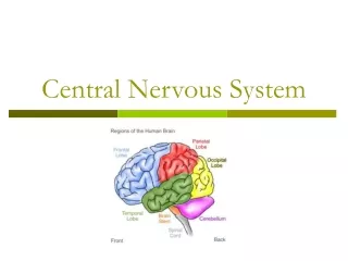
The Brain • Structure of the cerebrum • Cerebral cortex • Largest and uppermost division of the brain; consists of right and left cerebral hemispheres; each hemisphere is divided into five lobes : • Frontal lobe • Parietal lobe • Temporal lobe • Occipital lobe • Insula (island of Riel)
The Brain • Cerebral cortex (cont.) • Cerebral cortex—outer surface made up of six layers of gray matter • Gyri—convolutions; some are named: precentral gyrus, postcentral gyrus, cingulate gyrus, and hippocampal gyrus • Sulci—shallow grooves
The Brain • Cerebral cortex (cont.) • Fissures—deeper grooves, divide each cerebral hemisphere into lobes; four prominent cerebral fissures • Longitudinal fissure—deepest fissure; divides cerebrum into two hemispheres • Central sulcus (fissure of Rolando)—groove between frontal and parietal lobes • Lateral fissure (fissure of Sylvius) —groove between temporal lobe below and parietal lobes above; island of Reil lies deep in lateral fissure • Parietooccipital fissure—groove that separates occipital lobe from parietal lobes
The Brain • Functions of the cerebral cortex • Functional areas of the cortex—certain areas of the cerebral cortex predominantly engage in one particular function • Postcentralgyrus—mainly general somatic sensory area; receives impulses from receptors activated by heat, cold, and touch stimuli • Precentralgyrus—chiefly somatic motor area; impulses from neurons in this area descend over motor tracts and stimulate skeletal muscles • Transverse gyrus—primary auditory area • Occipital lobe—primary visual areas
The Brain • Functions of the cerebral cortex (cont.) • Sensory functions of the cortex • Somatic senses—sensations of touch, pressure, temperature, proprioception, and similar perceptions that require complex sensory organs • Cortex contains a “somatic sensory map” of the body • Information sent to primary sensory areas is relayed to sensory association areas, as well as to other parts of the brain • The sensory information is compared and evaluated, and the cortex integrates separate bits of information into whole perceptions
The Brain • Motor functions of the cortex • For normal movements to occur, many parts of the nervous system must function • Precentral gyrus—primary somatic motor area; controls individual muscles • Secondary motor area—in the gyrus immediately anterior to the precentral gyrus; activates groups of muscles simultaneously
The Brain • Integrative functions of the cortex • Consciousness State of awareness of one’s self, one’s environment, and other beings • Depends on excitation of cortical neurons by impulses conducted to them by the reticular activating system • There are two current concepts about the reticular activating system: • Functions as the arousal system for the cerebral cortex • Its functioning is crucial for maintaining consciousness • Language • Ability to speak and write words and ability to understand spoken and written words • Speech centers—areas in the frontal, parietal, and temporal lobes • Left cerebral hemisphere contains speech centers in approximately 90% of the population; in the remaining 10%, contained in either the right hemisphere or both • Aphasias—caused by lesions in speech centers
The Brain • Integrative functions of the cortex (cont.) • Emotions • Subjective experiencing and objective expressing of emotions involve functioning of the limbic system • Limbic system—also known as the “emotional brain” • Most structures of limbic system lie on the medial surface of the cerebrum; they are the cingulate gyrus and the hippocampus • Have primary connections with other parts of the brain, such as thalamus, fornix, septal nuclei, amygdaloid nucleus, and hypothalamus • Memory • One of the major human mental activities • Cortex is capable of storing and retrieving both short- and long-term memory • Temporal, parietal, and occipital lobes are among the areas responsible for short- and long-term memory • Engrams—structural traces in the cerebral cortex that comprise long-term memories • Cerebrum’s limbic system plays a key role in memory
The Brain • Hemisphericity—specialization of cerebral hemispheres • Right and left hemispheres of the cerebrum specialize in different functions; however, both sides of a normal person’s brain communicate with each other to accomplish complex functions • Left hemisphere is responsible for the following: • Language functions • Dominating control of certain hand movements • Right hemisphere is responsible for the following: • Perception of certain kinds of auditory material • Tactual perception • Perceiving and visualizing spatial relationships
Cycle of Life: Central Nervous System • The development and degeneration of the central nervous system is the most obvious functional change over the life span • Development of the brain and spinal cord begins in the womb • Lack of development in the newborn is evidenced by lack of complex integrative functions • Language • Complex memory • Comprehension of spatial relationships • Complex motor skills
Cycle of Life: Central Nervous System • Complex functions develop by adulthood • Late adulthood—tissues degenerate • Profound degeneration—unable to perform complex functions • Milder degeneration—temporary memory lapse or difficulty with complex motor tasks
The Big Picture: The Central Nervous System and the Whole Body • Central nervous system—ultimate regulator of the body; essential to survival • Able to integrate bits of information from all over the body, make sense of them, and make decisions