Download
1 / 58
910 likes | 3.39k Views
Antibody-Mediated Cytotoxicity ( Type II)Hypersensitivity 3 Mechanisms. Video clips . IgE mediated type I allergy ( http://www.healthcentral.com/allergy/video-44016-47.html ) MAC formation http://faculty.ccbcmd.edu/courses/bio141/lecguide/unit2/bacpath/mac_flash.html.

E N D
Antibody-Mediated Cytotoxicity (Type II)Hypersensitivity 3 Mechanisms
Video clips • IgE mediated type I allergy (http://www.healthcentral.com/allergy/video-44016-47.html) • MAC formation http://faculty.ccbcmd.edu/courses/bio141/lecguide/unit2/bacpath/mac_flash.html
Type II Hypersensitivity • Type II hypersensitivity involves IgG or IgM induced damage to self cells (Cell-surface or Matrix Antigen) • Either IgG or IgM is made • against normal self antigens- failure in immune tolerance • or a foreign antigenresembling some molecule on the surface of host cells enters the body and IgG or IgM made against that antigen then cross reacts with the host cell • Antibodies against drugs • Immune Processes involved: • Classical Complement Pathway • Phagocytosis via FcR and Complement receptor • ADCC via NK cells or eosinophils • Many autoimmune diseases result from type II hypersensitivity generated by autoantibodies Immunreaktionen der Haut
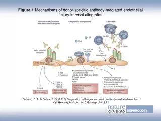
Ab can activate the complement system, creating pores in the membrane of a foreign cell Activation of the classical complement pathway causing MAC lysisof the cells
Abcan serve as an opsonin, enabling phagocytic cells with Fc or C3b receptors to phagocytose Opsonizationof the host cells whereby phagocytes stick to host cells by way of IgG, C3b, or C4b and discharge their lysosomes
It can mediate cell destruction by antibody dependent cell-mediated cytotoxicity (ADCC) whereby NK cells attach to the Fc portion of the antibodies The NK cell then release pore-forming proteins called perforins and proteolytic enzymes called granzymes Granzymes pass through the pores and activate the enzymes that lead to apoptosis of the infected cell by means of destruction of its structural cytoskeleton proteins and by chromosomal degradation
Examples of type II hypersensitivity • AB and Rh blood group reactions • autoimmune diseases such as: • rheumatic fever where antibodies result in joint and heart valve damage • idiopathic thrombocytopenia purpurawhere antibodies result in the destruction of platelets • myasthenia gravis where antibodies bind to the acetylcholine receptors on muscle cells causing faulty enervation of muscles • Goodpasture's syndrome where antibodies lead to destruction of cells in the kidney • multiple sclerosis where antibodies are made against the oligodendroglial cells that make myelin, the protein that forms the myelin sheath that insulates the nerve fiber of neurons in the brain and spinal cord; and some drug reactions • Type II hypersensitivity also participates in early transplant rejections
Transfusion Reactions Are Type II Reactions • Proteins and glycoproteins on the membrane of RBCs are encoded by different genes (with different alleles) • One allelic form of a blood-group Ag can recognize other allelic forms as foreign and mount an antibody response • Ab ( Isohaemagglutinins) IgMcan be induced by natural exposure to similar antigenic determinants on microorganisms ( normal flora) of gut • This is the case with the ABO blood-group Ags
Clinical manifestations in association with hemolytic anemia linked to blood transfusions • May occur due to massive hemolysis due to antibody and complement system • Immediate (ABO) IgM • Late IgG (2-6 days) • Symptoms include fever, low hemoglobin, increased bilirubin, mild jaundice, and anemia • Free hemoglobin is usually not detected in the plasma or urine in these reactions because RBC destruction occurs in extravascular sites
Antibodies to other blood-group AgsIgG class • May result from repeated blood transfusions because of minor allelic differences in these antigens • Delayed hemolytic transfusion reaction which may develop in 2 and 6 days reflecting the secondary nature of these reactions • The transfused blood induces clonal selection and production of IgG against a variety of blood-group membrane antigens • most commonly Rh, Kidd, Kell, and Duffy • The predominant isotype involved in these reactions is IgG- less effective than IgM in activating complement • many of the transfused cells are destroyed at extravascular sites • by agglutination • opsonization • and subsequent phagocytosis by macrophages
Type II HypersensitivityAntibody-Complement Dependent Mediated Lysis Example: Autoimmune Hemolytic Anemia Immunreaktionen der Haut
Reactions – immediate(hrs)with ABO incompatibilities • Complement- mediated lysis triggered by IgM • Free Hb in plasma; filtered through the kidneys, hemoglobinuria • Some Hbbilirubin, (toxic at high levels) • fever, chills, nausea, clotting within blood vessels, pain in the lower back, • Treatment- Stop transfusion , maintain urine flow with a diuretic, otherwise Hb in kidney acute tubular necrosis
Erythroblastosisfetalis, • Fetus- Rh+; Mother Rh- • Maternal IgGspecific for fetal blood-group Ags cross the placenta and destroy fetal RBCs . CF minor, serious, or lethal.. • During 1st pregnancy, fetal RBCs separated from the mother’s by trophoblast. • Delivery, separation of placenta from uterine wall fetal blood mother’s circulation. Rh-specific plasma cells and memory B cells in the mother. • Secreted IgMclears Rh+ fetal red cells from mother’s circulation, but memory cells remain, a threat to any subsequent pregnancy with an Rh+ fetus. • Activation of these memory cells in a subsequent pregnancy results in the formation of IgG anti-Rh antibodies, which cross the placenta and damage the fetal RBC .
…Cont • Severe anemia and brain damage due to lipid- soluble bilirubin • Rhogam antibodies given 24-48 hours after the birth, bind with fetal RBCs
ABO blood-group incompatibilityIn Newborn • (65%) of hemolytic disease of the newborn have minor consequences; caused by ABO blood-group incompatibility • Type A or B fetuses with Type O mothers IgGto A or B - natural exposure or through exposure to fetal blood-group in successive pregnancies. • Usually fetal anemia is mild; Slight elevation of bilirubin, • Blood-exchange transfusion may be required in infants. • Low levels of UV light is enough to break down bilirubin.
Diagnosis • Rising titer of Ab to Rh Ag in mother in pregnancy • Maternal IgG on the surface of fetal RBC are detected by Coombs test.
Treatment • Severe reaction- intrauterine blood-exchange transfusion. • Less severe cases- blood-exchange transfusion after birth, UV light • Mother Tt during pregnancy by plasmapheresis
Type II Response With Antibiotics Drug such as: Pencillin Cephaliosporin Streptomycin May act as hapten- protein complexes(Ag) adsorb on RBCs Ab attaches to Ag (adsorbed drug) induces complement activation lysis progressive anemia
Immune Complex–Mediated(Type III) Hypersensitivity • Complexingof Ag with Ab facilitates the clearance of antigen by phagocytic cells. • Large amounts of immune complexes can lead to tissue-damaging type III hypersensitive reactions; • The severity may be dependent on the quantity & distribution of immune complexes • Localized inflammation if at the site of antigen entry • Widespread inflammation where ever complexes deposits through blood
Type III Hypersensitivity • “Immune complex disease” • Soluble Ag/IgG or IgM • high titers of each required • Immune processes involved: • classical complement pathway • phagocytic cells Immunreaktionen der Haut
Type-III Hypersensitivity: Immune Complex Animation: Large quantities of soluble antigen-antibody complexes form in the blood and are not completely removed by macrophages. These antigen-antibody complexes lodge in the capillaries between the endothelial cells and the basement membrane. The antigen-antibody complexes activate the classical complement pathway and complement proteins and antigen-antibody complexes attract leukocytes to the area. The leukocytes then discharge their killing agents and promote massive inflammation. This leads to tissue death and hemorrhage Immunreaktionen der Haut
Pathogenesis • Ag – Ab complexes +C3b as opsoninrecruitment of neutrophils(R for C3b)- tissue injury as granules release • C3a, C4a, and C5a (complement split products) • anaphylatoxin localized mast-cell degranulation • C3a, C5a and C5b67 are also chemotactic factor for neutrophils • Damage caused due to innocent bystander lysis • Activation of C aggregation of plateletsrelease of clotting factors formation of microthrombi
Sites of Complex Deposition Site Outcome glomeruli glomerulonephritis blood vessel wall arteritis synovial membrane arthritis skin rash Note: Ab responsible for immune complexes may be generate at a site distant from the point of deposition. Immunreaktionen der Haut
Arthus Reaction- localized type III hypersensitivity • Inj of Ag ID or S/C who has high levels of circulating Ab specific for that Ag localized immune complexes acute Arthus reaction within 4–8 h • Microscopic- neutrophils adhering to the vascular endothelium migrate to tissues towards complexes • Insect bite( sensitive individual) rapid localized type I reaction 4–8 h later Arthus reaction with pronounced erythema and edema • Intrapulmonary Arthus-type reactions induced by bacterial spores, fungi, or dried fecal proteins pneumonitis or alveolitis • Disease names reflecting source of Ag • “farmer’s lung” inhalation of thermophilicactinomycetes from moldy hay, • “pigeon fancier’s disease” from inhalation of serum protein in dust derived from dried pigeon feces
Arthus Reaction • As the reaction develops, localized tissue and vascular damage results in an accumulation of fluid (edema) and red blood cells (erythema) at the site • The severity of the reaction can vary from mild swelling and redness to tissue necrosis
Type III Generalized Reactions -Serum Sickness • Large amounts of Ag enter bloodstream & bind to Abcirculating immune complexes • If Ag in excess, small complexes form; not easily cleared by the phagocyticcells tissue-damaging at various sites of deposition • Serum sickness - antitoxins containing foreign serum • E.g. horse antitetanus and antidiptheria serum • Symptoms appear within days and weaks after exposure • These symptoms include fever, weakness, generalized vasculitis (rashes) with edema and erythema, lymphadenopathy, arthritis, and sometimes glomerulonephritis
Serum sickness Immunreaktionen der Haut
Serum Sickness • The precise manifestations of serum sickness depend on • Quantity of immune complexes • Size of complexes( determine the site of deposition) • Complexes accumulate in tissues where filtration of plasma occurs. This explains the high incidence of • glomerulonephritis(complex deposition in the kidney) • vasculitis(deposition in the arteries) and • arthritis (deposition in the synovial joints)
Other diseases caused due to the formation of Immune Complexes • 1. Autoimmunity (SLE, RA, Goodpasture’s ) • 2. Drug Reactions (Allergy to penicillin etc ) • 3.Infectious Diseases(Poststreptococcalglomerulonephritis, Meningitis, Hepatitis, Mononucleosis, Malaria, Trypanosomiasis
Type IV or Delayed-TypeHypersensitivity (DTH) • Subpopulations of activated TH cells encounter Ag,secrete cytokines localized inflammatory reaction called (DTH) • Characterized by large influxes of nonspecific inflammatory cells, in particular, macrophages • 1890 by Robert Koch, “tuberculin reaction” Later named DTH
Examples of Type IV Hypersensitivity Immunreaktionen der Haut
TH1 Influence of Immune Response • The hallmarks of a type IV reaction are • the delay in time required for the reaction to develop • recruitment of macrophages as opposed to neutrophils (type III reaction) Immunreaktionender Haut
TH1-mediated Type IV Hypersensitivity • Initial sensitization phase is 1-2 weeks after the primary contact • Primary APCs are: • Macrophages • Langerhans cells • MHCII+- Vascular endothelial cells • During this phase • Activation of TH cells (TH1 and CTLs) • Clonal expansion of TH cells
Pathways of Cytotoxicity utilized by CTL’s • Effector phase: • Starts after 24hrs, peaks at 48-72 hrs • Initiates due to recruitment of non-specific immune cells due to TH1 (5%) mediated cytokines • Denovo synthesis of macrophages from blood monocytes • DTH response is important for defense against intracellular parasites and bacteria (phagocytic cells, lytic enzymes) • However prolonged DTH can be damaging to the host
Contact Dermatitis DTH due to either TH1 or CTL mediated hypersensitivity Immunreaktionen der Haut
DTH is not always detrimental • Granuloma formation (fusion of continuously activated macrophages lead to giant multinucleated cells) • These giant cells displace the normal tissue cells, forming palpable nodules • granuloma-type lesion called a tubercle • e.g. M. Tuberculosis, activated macrophages wall off bacterium in lungs • and release high concentrations of lytic enzymes, which destroy bacterium and perhaps the surrounding tissue - extensive tissue necrosis
Development of delayed-type hypersensitivity reaction • after a second exposure to poison oak • Cytokines such as IFN-, macrophage-chemotactic factor (MCF), and migration-inhibition factor (MIF) released from sensitized TH1 cells mediate this reaction • Tissue damage results from lytic enzymes released from activated macrophages
Patch test This test is used to diagnose delayed allergic reactions such as Contact Dermatitis. It involves taping traces of various known contact allergens on the skin and keeping them there for 48 hours It can test for allergy to Rubber, Nickel, Lanolin, dyes, cosmetics, solvents, preservatives, and medication. Immunreaktionen der Haut
Neutrophils – first to appear, peaking 6h • The monocyte- 24 -48 h after antigen exposure • MCAF (monocytechemotactic and activating factor)- recruitment of macrophages • MIF (mirgration inhibitory factor)- inhibits migration of macrophages beyond DTH
IFN Importance • knockout mice for IFN when infected with an attenuated strain of Mycobacterium bovisknown as BCG (BacilleCalmetteGuérin), nearly all the animals died within 60 days, whereas wild-type mice survived • Macrophages from the IFN-knockout mice were shown • to have reduced levels of class II MHC molecules • ndof bactericidal metabolites such as • nitric oxide • superoxide anion
http://highered.mcgraw-hill.com/sites/0072507470/student_view0/chapter22/animation__delayed__type_iv__hypersensitivity.htmlhttp://highered.mcgraw-hill.com/sites/0072507470/student_view0/chapter22/animation__delayed__type_iv__hypersensitivity.html
Hypersensitive reactions on the basis of effector molecules rather than antigens
Contribution of environmental factors on allergy susceptibility
Environmental factors may interact with genetic susceptibility to cause allergic disease • environmental factors and genetic variation each account for about 50% • The prevalence of atopic allergic diseases- is increasing in economically advanced regions • Environmental changes- involves exposure to infectious diseases in early childhood • Change from traditional rural society meant less exposure to microbes • Changes in intestinal microbiota • idea that exposure to microorganisms is associated with allergy was first mooted in 1989- hygiene hypothesis
Hygiene hypothesis to counter- regulation hypothesis • The original proposition was that less hygienic environments → infections early in childhood → help to protect against the development of atopy and allergic asthma • mechanisms that skewed immune responses towards TH1 rather than TH2 cells & their associated cytokines → IgE production • A study in Venezuela showed that children treated for a prolonged period with antihelminthic agents had a higher prevalence of atopy than did untreated and heavily parasitized children