Download
1 / 62
740 likes | 1.83k Views
Acute Respiratory Distress Syndrome (ARDS). Prepared by Wajeeha Nabulsi, Ghassan Zakarni Supervised by Dr. Aidah Abu Elsoud Alkaissi An-Najah National University Faculty of Nursing. Another Names for ARDS. Da Nang Lung Transfusion Lung Post Perfusion Lung Shock Lung

E N D
Acute Respiratory Distress Syndrome (ARDS) Prepared by Wajeeha Nabulsi, Ghassan Zakarni Supervised by Dr. Aidah Abu Elsoud Alkaissi An-Najah National University Faculty of Nursing
Another Names for ARDS • Da Nang Lung • Transfusion Lung • Post Perfusion Lung • Shock Lung • Traumatic Wet Lung • Posttraumatic Failure • Posttraumatic Pulmonary Insufficiency • Wet lung • White Lung
HISTORICAL PERSPECTIVES • Described by William Osler in the 1800’s • Ashbaugh, Bigelow and Petty, Lancet – 1967 • 12 patients • pathology similar to hyaline membrane disease in neonates
ORIGINAL DEFINITION • Acute respiratory distress • Cyanosis refractory to oxygen therapy • Decreased lung compliance • Diffuse infiltrates on chest radiograph • Difficulties: • lacks specific criteria • controversy over incidence and mortality
REVISION OF DEFINITIONS • 1988: four-point lung injury score • Level of PEEP • PaO2 / FiO2 ratio • Static lung compliance • Degree of chest infiltrates
1994 CONSENSUS • Acute onset • may follow catastrophic event • Bilateral infiltrates on chest radiograph • PAWP < 18 mm Hg • Two categories: • Acute Lung Injury - PaO2/FiO2 ratio < 300 • ARDS - PaO2/FiO2 ratio < 200
INCITING FACTORS • Shock • Aspiration of gastric contents • Trauma • Infections • Inhalation of toxic gases and fumes • Drugs and poisons • Miscellaneous
STAGES • Acute, exudative phase • rapid onset of respiratory failure after trigger • diffuse alveolar damage with inflammatory cell infiltration • hyaline membrane formation • capillary injury • protein-rich edema fluid in alveoli • disruption of alveolar epithelium
STAGES • Subacute, Proliferative phase: • persistent hypoxemia • development of hypercarbia • fibrosing alveolitis • further decrease in pulmonary compliance • pulmonary hypertension
STAGES • nn • Chronic phase • obliteration of alveolar and bronchiolar spaces and pulmonary capillaries • Recovery phase • gradual resolution of hypoxemia • improved lung compliance • resolution of radiographic abnormalities
MORTALITY • 40-60%Deaths due to: • multi-organ failure • Sepsis • Mortality may be decreasing in recent years • better ventilatory strategies • earlier diagnosis and treatment
PATHOGENESIS • Inciting event • Inflammatory mediators • Damage to microvascular endothelium • Damage to alveolar epithelium • Increased alveolar permeability results in alveolar edema fluid accumulation
PATHOGENESIS • Target organ injury from host’s inflammatory response and uncontrolled liberation of inflammatory mediators • Localized manifestation of SIRS • Neutrophils and macrophages play major roles • Complement activation • Cytokines: TNF-a, IL-1b, IL-6 • Platelet activation factor • Eicosanoids: prostacyclin, leukotrienes, thromboxane • Free radicals • Nitric oxide
PATHOPHYSIOLOGY • Abnormalities of gas exchange • Oxygen delivery and consumption • Cardiopulmonary interactions • Multiple organ involvement
ABNORMALITIES OF GAS EXCHANGE • Hypoxemia: HALLMARK of ARDS • Increased capillary permeability • Interstitial and alveolar exudate • Surfactant damage • Decreased FRC • Diffusion defect and right to left shunt
ARDS • Is defined as a distinct form of acute respiratory failure that results from diffuse pulmonary injury of various causes • characterized by: - diffuse alveolar - capillary wall injury - increased alveolar- capillary permiability - noncardiogenic pulmonary edema - hyaline membrane formation, and atelectasis
Pathophysiology • The physiologic reaction of all body tissues sometimes results in pathologic changes in the lung • Asystemic insults causes low tissue perfusion and cellular hypoxia • Consequently, peripheral tissues are deprived of essential nutrients, and intracellular metabolic derangements result
Pathophysiology • Certain chemical factors such as prostaglandins, clotting factors, lysosomal enzymes, activated complement, or histamine are released into the systemic circulation • Prostaglandin contribute to vasodilation, capillary permeability, pain and fever, which accompany cell injury • Changes in the vessel walls and disturbances in blood flow increase platelet function, causing adhesiveness and aggregation
Pathophysiology • Lysosomal enzymes from neutrophils increase vascular permeability and cause tissue damage • Histamine is released from platelets, mast cell, and basophils and cause arterial vasodilation and enhanced permiability of capillaries and venules
Pathophysiology • Neutrophils and other inflammatory mediators can thus gain access to the lung parenchyma and carry on the inflammatory process • The inflammation then produces the lung injury, Severe ventilation-perfusion mismatching occurs • Alveolar collaps of the inflammatory infiltrate, blood fluid and surfactant dysfunction
Pathophysiology • The lung compliance becomes markedly decreased (stiff lung) • The blood returning to the lung for gas exchange is pumped through the nonventilated, nonfunctioning areas of the lung, causing a shunt to develop • The blood interfacing with nonfunctioning alveoli and gas exchange is markedly impaired, resulting in severe refractory hypoxemia
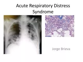
Clinical Features of ARDS • The earliest clinical signs of ARDS include tachypnea and progressive hypoxemia • Within 24 hours, the chest x-ray begins to reveal bilateral pulmonary infiltrates • Progression to mechanical ventilation often occurs in the first 48 hours of the illness
Common conditions that predispose to ARDS • Aspiration (gastric secretion, drowning, hydrocarbons) • Intracranial hypertension • Haemetologic disorders (disseminated intravascular coagulopathy (DIC) , Massive transfusion of blood products, cardiopulmonary bypass • Prolonged inhalation of high concentration of oxygen, smoke, or corrosive substances • Shock (any cause) • Catheter sepsis, drugs
Common conditions that predispose to ARDS • Localized infection (bacterial, fungal, viral pneumonias), • Trauma (pulmonary contusion, multiple fracture, head injury) • Major surgery • Metabolic disorders (Pancreatitis, uremia) • Urosepsis, • amniotic fluid embolism • Long bone fracture • Fat or air embolism • Systemic sepsis
Implementation and evaluation1. Fluid therapy • Hypoalbuminemic patients should receive coloids whereas all other patients should receive crystalloid fluids to decrease the pulmonary congestion • The patient´s pulmonary capillay wedge pressure (PCWP) is kept as low as possible as long as the cardiac output and tissue perfusion can be maintained at normal levels • Maintenance of the PCWP at 10-15 mm Hg provides adequate, but not excessive intravascular volumes
Maintaining tissue oxygenation • The fractional concentration of inspired oxygen (FiO2) should be kept at 50% or lower to minimize the risk of oxygen toxicity • An SaO2 above 90% should be sufficient to maintain oxygen delivery to peripheral tissues • If the FiO2 cannot be reduced to below 60% external PEEP is added to help reduce the FiO2 to nontoxic levels
2. Maintaining Tissue Oxygenation • The goal of O2 therapy is to administer the lowest possible oxygen concentration to sustain a mixed venous oxygen greater than 40 mm Hg • Positive end expiratory pressure (PEEP) is indicated for use in patients who are being ventilated mechanically with high FiO2 (>0.50) and who have a PaO2 of less than 65 mm Hg • The purpose of PEEP in ARDS is to minimize alveolar collapse and small airway closure and reduce interstitial edema and total extravascular lung water
2. Maintaining Tissue Oxygenation • Initial levels of PEEP should be in the range of 5-10 cm H2O • Small increments of PEEP are added until the optimal level is reached • The best meaurement available for evaluating tissue oxygenation at the bedside are systemic oxygen uptake (VO2 [oxygenconsumption]), venous lactate level, and gastric intramucosal PH ( measure directly by gastric tonometry)
2. Maintaining Tissue Oxygenation • Tissue oxygenation is considered to be inadequate if whole body VO2 is less than 100 ml/minute/m2 , venous lactate is greater than 4 mmol/L, or gastric intramucosal PH is less than 7.32
3. Drug Therpy • Morphine for sedating mechanically ventilated pat, who are restless, fearful and experiencing tachypnea • Pancuronium bromide (pavulon) neuroblocking agent to paralyze completely the voluntary respirations of the patient • Possible sedatives are: lorazepam (ativan), midazolam (versd), haloperidol (haldol), propofol (diprivan), and short-acting barbiturates.
4. Preventing Iatrogenic Injury Ventilator management • There is now considerable evidence indicating that the large tidal volume used during conventional mechanical ventilation (10 to 15 ml/kg) can damage the lungs • The pathologic changes in ARDS are not distributed uniformly throughout the lungs
4. Preventing Iatrogenic Injury Ventilator management • Recognition of the risk of lung injury at high inflation volumes and pressures had led to an alternative strategy where peak inspiratory pressures are kept below 35 cm H2O by using tidal volume of 7-10 ml/kg • According to this strategy, mechanical ventilation is started at inflation volumes of 10 ml/kg • If the resulting peak inspiratory pressure (PIP) is above 35 cm H2O, the inflation volume is reduced in increments of 2 ml/kg until PIP falls below 35 cm H2O
5. Reducing Lung Water • The two measures that are advocated for reducing lung water are diuretics and PEEP • Unfortunately , neither measure is likely to be effective in ARDS • The application of PEEP does not reduce extravasascular lung water in ARDS
5. Reducing Lung Water Diuretics • The use of diuretics to minimize or reduce fluid overload seems a more reasonable measure, but only when renal water excretion is impaired (otherwise the best way to prevent fluid overload is to maintain an adequate cardiac output)
Positive End-Expiratory Pressure • In fact, high levels of PEEP can actually increase lungwater • This latter effect may be the result of alveolar overdistension, or may be the result of PEEP-induced impairment of lymphatic drainage from the lungs
If there is evidence for impaired tissue oxygenation, the sequence of managementCardiac Output • If the cardiac output is inadequate (e.g. A cardiac index below 3L/min/m2 and CVP or wedge pressures are not elevated, volume infusion is indicated • If volume infusion is not indicated, dobutamine is used to augment the cardiac output • Dopamine should be avoided because of its propensity to constrict pulmonary vein
If there is evidence for impaired tissue oxygenation, the sequence of managementBlood Transfusion • Transfusion is often recommended to keep the Hb above 10 g/dL. • In fact, given the propensity for blood transfusion to cause ARDS • It seems wise to avoid transfusing blood products in ARDS • If there is no evidence of inadequate tissue oxygenation, there is no need to correct anemia
A brief summary of the available studies- Steroids • High-dose methylprednisolone (30 mg/ kg) I.V every 6 hours for 4 doses) given to patients within 24 hours of the diagnosis of ARDS has not improved outcome or reduced mortality • In fact, one study showed a higher mortality associated with steroid therapy in ARDS (Bone 1987) • Secondary infection are increased in patients receiving high dose methyl-prednisolone for ARDS (Bone 1987)
A brief summary of the available studies- Steroids • High-dose methylprednisolone (2-3 mg/kg/day) given to 25 patients with late ARDS (2 weeks duration) resulted in a beneficial response in 21 patients and an 86% survival in the responders (Meduri 1994) • This study suggests a possible role for steroids late in the course of ARDS, but corroborative evidence is required
Specific Therapies1. Surfactant • Aerosolized surfactant has proven effective in improving outcomes in the neonatal form of respiratory distress syndrome, but it has not met with similar success in adults with ARDS (Anzueto 1996)
Specific Therapies2. Antioxidant • Neutrophil-mediated tissue injury may play an important role in the pathogenesis of ARDS, it is no surprise that there is conciderable interest in the possible role of antioxidants as a specific theray for ARDS
3. Nitric oxide • Nitric oxide can improve oxygenation and reduce pulmonary artery pressures in ARDS, mortality is unchanges (Lunn 1995) • Nitric oxide is a pulmonary vasodilator, which inhaled crosses the alveolar membrane and acts locally on the pulmonary vasculature, dilating vessels and increasing blood flow
3. Nitric oxide • Has the effect of improving ventilation/perfusion (V/Q) matching and therefore gas exchange as the blood flow is only increased in the ventilated areas • as soon as it enters the blood, nitric oxide is bound to haemoglobin and has no further systemic (i.e. Hypotensive) effect
Specific Therapies • Numerous pharmacological RX are underinvestigation to stop the cascade of events leading to ARDS • Neutrrophil inhibitors • Interleukin-1 receptor antagonist • Pulmonary specific vasodilator • Surfactant replacement therapy • Antisepsis agents
prone position Study shows that prone positioning significantly improves oxygenation in about 65% of patients. This allows nurses to reduce the percentage of inspired oxygen and positive end-expiratory pressure.
Prone positioning • Prone therapy assists pts with ARDS by reducing the ventilation\perfusion mismatch
Kinetic Therapy™ Can help improve clinical outcomes and lower overall cost of care. By continuously rotating critically ill patients from side-to-side to at least 40°, Kinetic Therapy™ helps prevent and treat pulmonary complications as ARDS