Download
1 / 7
70 likes | 117 Views
This study compares the amount of H2O2 generated by glucose oxidase and the impact of PARP1 inhibition on mitochondrial DNA repair. The figure displays fluorescence increase in response to varying enzyme concentrations and H2O2 levels, as well as expression levels of DNA repair enzymes. Data from independent experiments show repair rates in untreated and inhibitor-treated cells. Localization of PARP1 in mitochondria and its interaction with repair enzymes are also examined.

E N D
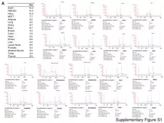
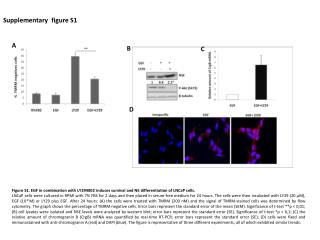
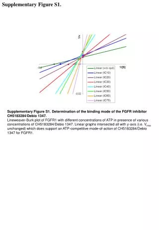
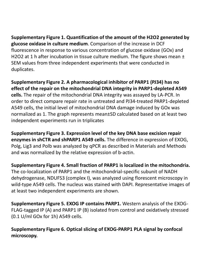
Supplementary Figure 1. Quantification of the amount of the H2O2 generated by glucose oxidase in culture medium. Comparison of the increase in DCF fluorescence in response to various concentration of glucose oxidase (GOx) and H2O2 at 1 h after incubation in tissue culture medium. The figure shows mean ± SEM values from three independent experiments that were conducted in duplicates. Supplementary Figure 2. A pharmacological inhibitor of PARP1 (PJ34) has no effect of the repair on the mitochondrial DNA integrity in PARP1-depleted A549 cells. The repair of the mitochondrial DNA integrity was assayed by LA-PCR. In order to direct compare repair rate in untreated and PJ34-treated PARP1-depleted A549 cells, the initial level of mitochondrial DNA damage induced by GOx was normalized as 1. The graph represents mean±SD calculated based on at least two independent experiments run in triplicates Supplementary Figure 3. Expression level of the key DNA base excision repair enzymes in shCTR and shPARP1 A549 cells. The difference in expression of EXOG, Polg, Lig3 and Polb was analyzed by qPCR as described in Materials and Methods and was normalized by the relative expression of b-actin. Supplementary Figure 4. Small fraction of PARP1 is localized in the mitochondria. The co-localization of PARP1 and the mitochondrial-specific subunit of NADH dehydrogenase, NDUFS3 (complex I), was analyzed using florescent microscopy in wild-type A549 cells. The nucleus was stained with DAPI. Representative images of at least two independent experiments are shown. Supplementary Figure 5. EXOG IP contains PARP1. Western analysis of the EXOG-FLAG-tagged IP (A) and PARP1 IP (B) isolated from control and oxidatively stressed (0.1 U/ml GOx for 1h) A549 cells. Supplementary Figure 6. Optical slicing of EXOG-PARP1 PLA signal by confocal microscopy.
Supplementary Figure S1 30uM H2O2 100uM H2O2 300uM H2O2 0.01 U/ml GOx 0.03 U/ml GOx 0.003 U/ml GOx
Supplementary Figure S4 DAPI PARP1 merge NDUFS3
Supplementary Figure S5 A. IP input EXOG-FLAG EXOG-FLAG EXOG-FLAG vector vector vector WB: EXOG FLAG PARP1 - - + + GOx B. IP: PARP1 WB: PARP1 EXOG - + GOx