Download
1 / 57
580 likes | 784 Views
EM Clerkship: Intro to Trauma Care. Objectives. Trauma Epidemiology Trauma Primary Survey Life-threats during primary survey Identification Treatment Traumatic Brain Injury Spine protection/clearance. Epidemiology. 30K deaths annually in U.S. Leading cause of death 1-44 y.o.

E N D
Objectives • Trauma Epidemiology • Trauma Primary Survey • Life-threats during primary survey • Identification • Treatment • Traumatic Brain Injury • Spine protection/clearance
Epidemiology • 30K deaths annually in U.S. • Leading cause of death 1-44 y.o. • Lost years > heart disease, stroke, & cancer combined • In next 30 minutes • 6 people will die • 1000 people will have a disabling injury • $24 million will be spent on these patients
Clinical Case 19 yo male in highway speed motor vehicle crash, ejected from vehicle, with decreased level of consciousness at scene. Arriving in ED in 5 minutes. Vital Signs: P 125 bpm, BP 85/50, RR 35, T 36 C. Prepare for this patient now . . .
Advanced Trauma Life Support(ATLS) • Primary Survey • Resuscitation • Secondary Survey • Definitive Care
Trauma: Primary Survey • Rapid assessment of patient with trauma • Immediate recognition of and intervention for life-threatening injuries • Airway • Breathing • Circulation • Disability • Exposure/Environment
Trauma Care • ABCs are the core of caring for a “sick” trauma patient • Revisit and Reassess • Does the patient have an A problem, a B problem, or a C problem? • ADDRESS THESE IMMEDIATELY!
Clinical Case 19 yo male s/p MVC, arrives to ED with Medics. Vital Signs: P 125 bpm, BP 85/50, RR 35, T 36 C. How do you assess and manage his airway?
Airway • Look • Fractures, foreign bodies, ongoing bleeding • Signs of neck or upper chest trauma • COLLAR: CERVICAL SPINE PROTECTION • Listen • Can the patient talk easily? • Stridor, changes in voice quality, snoring, gurgling • Feel • Air exchange
Treatment: Airway Obstruction • Jaw thrust • Cervical spine immobilization • Adjunct airway equipment • Oropharyngeal airway • Endotracheal Intubation • Surgical airway
Clinical Case 19 yo male s/p MVC You intubated on arrival for airway control. Vital Signs: P 125 bpm, BP 85/50, RR on vent, T 36 C. How do you assess and manage his breathing?
Breathing • Look • Symmetric chest rise • Depth and rate of respirations • Listen • Symmetric breath sounds • Feel • Crepitus • Fractures/deformity
Clinical Case 19 yo male s/p MVC A:You intubated on arrival for airway control. B: Chest tube placed on R for diminished BS and crepitus. Vital Signs: P 125 bpm, BP 85/50, RR on vent, T 36 C. How do you assess and manage his circulation?
Circulation • Look • Vital signs—HR, BP • Skin color • IV access: What is adequate? • Feel • Skin • Pulses • Address • Direct pressure to active bleeding • IV fluid: What is your order? • They don’t respond to fluid bolus. What next?
Clinical Case 19 yo male s/p MVC A: Intubated B: Chest tube C: 2 x 16g IV in ACs, 2L LR bolus Vital Signs: P 105 bpm, BP 97/57, RR on vent, T 36 C. How do you assess and manage his disability?
Disability • Assess • Mental status • GCS • Pupillary response • Moving extremities?
EYES Spontaneous (4) To Speech (3) To Pain (2) Closed (1) VERBAL Oriented (5) Confused (4) Inappropriate (3) Incomprehensible (2) None (1) If intubated, 1T MOTOR Obeys (6) Localizes (5) Withdraws (4) Flexion (3) Extension (2) None (1) Glasgow Coma Score Mild TBI: 13-15 Moderate: 9-12 Severe: ≤ 8
Clinical Case 19 yo male s/p MVC A: Intubated B: Chest tube C: 2 x 16g IV in ACs, 2L LR bolus D: GCS 8T, withdraws toes to pain Vital Signs: P 105 bpm, BP 97/57, RR on vent, T 35 C. How do you expose and manage environment?
Exposure/Environment • Look • Expose the patient • Cut off clothes with shears • Roll, look for injuries • Environment • Avoid hypothermia • Warm blankets and fluids
Adjuncts to Primary Survey • Safety net • What is “adequate” IV access? • Labs • Portable X-rays (“trauma series”) • Lateral C-spine • CXR • AP Pelvis • FAST exam • Early surgical consultation as indicated • Foley, NG tube, etc.
Trauma Series • What can the trauma series tell us?
Life-threatening Chest Injuries: What kind of problem? • A: Airway Obstruction • B: Tension Pneumothorax • B: Open Pneumothorax • B: Flail chest • B/C: Massive Hemothorax • C: Cardiac Tamponade
NAME the DIAGNOSIS • No air flow through mouth, can not ventilate the patient • Compromised vital signs, decreased breath sounds on right, neck veins distended, trachea deviated to left • Compromised vital signs, neck veins distended, heart tones distant, low voltages on ECG
NAME the DIAGNOSIS • Compromised vital signs, decreased breath sounds on right, neck veins flat, dull percussion on right • Respiratory distress, compromised ventilation, bony crepitance of chest wall • Respiratory distress, compromised ventilation, “gurgling” hole in chest wall
Tension Pneumothorax http://www.med.yale.edu/intmed/cardio/imaging/cases/pneumothorax_tension http://cfs2.tistory.com/
Tension Pneumothorax • Physical exam findings • Vital sign changes • Distended neck veins • Unilateral absence of breath sounds • Hyperresonant to percussion • Tracheal deviation AWAY from affected side • Clinical diagnosis • “One-way valve” air leak • Air gets into pleural space, can’t get out • Pressure decreases venous return • WORSENED by positive pressure ventilation
Treatment: Tension Pneumothorax • Needle thoracostomy • 14 gauge needle • 2nd intercostal space • Midclavicular line • OVER the rib • Temporizing measure until chest tube placed http://www.aic.cuhk.edu.hk/web8/Hi%20res/Needle%20thoracostomy.jpg
Tube Thoracostomy Roberts: Clinical Procedures in Emergency Medicine, 4th ed.
Open Pnemothorax • AKA “Sucking Chest Wound” • Traumatic defect in chest wall • Thorax equilibrates with external pressure, resulting in INCREASED intrathoracic pressure
Treatment: Open Pneumothorax • Flutter valve dressing • Tape occlusive dressing on three sides • Air can exit wound, but doesn’t enter with inspiration • Do NOT seal all edges • TENSION PNEUMOTHORAX risk • Chest tube will be needed
Flail Chest • Two or more consecutive fractured ribs, each fractured in two or more places • A segment of the chest wall is not connected with the rest of the chest • Exam: Paradoxical chest wall movement, hypoxia, respiratory distress, crepitus • CXR: Fractures, pulmonary contusion • Treatment: Ventilation, oxygenation, ABG, analgesia, consider intubation
Flail Chest http://www.itim.nsw.gov.au/images/Right_Flail1.jpg
Cardiac Tamponade • Can happen from blunt or penetrating injury • Blood in pericardium increases pressure, decreasing venous return and perfusion • Beck’s triad • Distended neck veins • Hypotension • Muffled heart tones • Pulsus paradoxus • Normal decrease in SBP during inspiration 10mm Hg • >10mm Hg decrease is sign of tamponade
Cardiac Tamponade • Treatment: pericardiocentesis or thoracotomy (with cardiac arrest)
Open Thoracotomy • Indications • Cardiac arrest from penetrating chest wound • Some patients with witnessed cardiac arrest following blunt chest trauma • Objectives • Release cardiac tamponade • Control hemorrhage • Cardiac massage • Repair cardiac wounds • Compress descending aorta
Open Thoracotomy • Considerations • Very low survival rate (1-9%) • Sharps/exposure risk to providers • Technique • Sterile prep • Left anterolateral approach, 4th-5th intercostal space from sternum to mid-axillary line • Incise with scalpel down through intercostals • Spread with rib spreader • Internal procedures as indicated
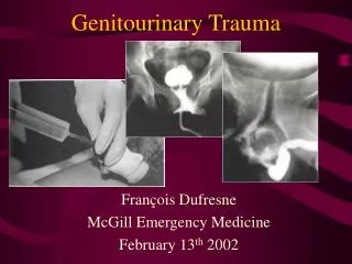
Aortic Injury • Majority die at scene • Can be a primary survey issue • Increased risk with rapid deceleration force (motor vehicle crash, high fall) • CXR findings • Widened mediastinum • Obliteration of aortic knob • Deviation of trachea to right • Left hemothorax • Needs angiography to diagnose (usually CT)
Aortic Injury http://www.surgery.ucsf.edu/eastbaytrauma/images/ctarrow.jpg
NEJM 2001; 344(8): 580. NAME THE THREE TYPES OF ICH SEEN HERE
HEAD INJURY • 50% of Trauma Deaths • 500,000 CHI hospitalized annually • Emergency Care • Always Consider a head injury in multi-trauma • GCS, and repeat examination • Neurosurgery consultation • Manage Increased ICP • Maintain pCO2 30-35 • Elevate HOB
CLINICAL QUESTION You are a senior medical student in the HMC ED. An unstable multi-trauma patient has arrived. Over the course of the patient’s ED care, during the “Secondary Survey” the Trauma Doctor asks you to place a nasogastric tube for stomach decompression. What findings on head & face examination would contraindicate nasal placement of this tube?
Trauma: Secondary Survey • Reevaluate ABCDEs • Careful head to toe evaluation of patient • High index of suspicion for other injuries
Definitive Care • Primary Survey • Address problems with ABCDE • STOP the bleeding if possible • Early consultation • Trauma surgeons • Neurosurgeons • Other specialists (ortho, ophtho, oto, etc.) • May include transfer to trauma center