Download
1 / 1
20 likes | 290 Views
A. B. APP. APP. AA. A. B. TC tórax plano axial (ventana mediastino). A) Valoración diámetro de la arteria pulmonar principal ( 29 mm). B) Valoración del receso pericárdico superior (flecha). AA: aorta ascendente. APP: Arteria pulmonar principal.

E N D
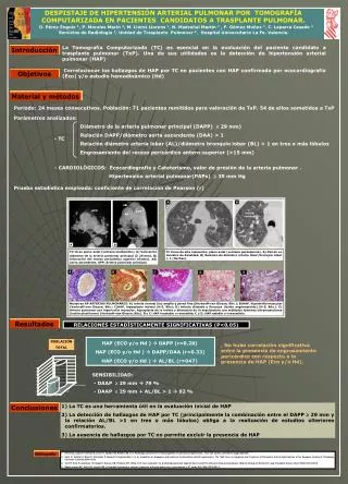
A B APP APP AA A B TC tórax plano axial (ventana mediastino). A) Valoración diámetro de la arteria pulmonar principal ( 29 mm). B) Valoración del receso pericárdico superior (flecha). AA: aorta ascendente. APP: Arteria pulmonar principal. TC tórax de alta resolución, plano axial (ventana parénquima). A) Patrón en mosaico de densidad. B) Relación de diámetro arteria lobar/bronquio lobar > 1 (flechas). A B C D E Muestras AP ARTERIAS PULMONARES: A) arteria normal (luz amplia y pared fina)(Verhoeff-van Gieson, 80x.); B)HAP, hipertrofia muscular (Verhoeff-van Gieson, 80x.; C)HAP, hiperplasia intimal (H-E. 80x); D) Arteria dilatada y flexuosa (lesión angiomatoide) (H-E, 80x.). E) Arteria pulmonar con hipertrofia muscular, hiperplasia de la íntima y alteración de la arquitectura con múltiples lúmenes intramusculares (lesión plexiforme) (Verhoeff-van Gieson, 80x). B y C: HAP inestable o reversible; C y D: HAP estable o irreversible. HAP (ECO y/o Hd ) DAPP (r=0.28) HAP (ECO y/o Hd ) DAPP/DAA (r=0.33) HAP (ECO y/o Hd ) AL/BL (r=047) DESPISTAJE DE HIPERTENSIÓN ARTERIAL PULMONAR POR TOMOGRAFÍA COMPUTARIZADA EN PACIENTES CANDIDATOS A TRASPLANTE PULMONAR. D. Pérez Enguix 1, P. Morales Marín 2, M. Lloret Llorens 1, N. Madroñal Martín 1 , F. Gómez Muñoz 1, C. Laparra Casado 1 Servicios de Radiología 1, Unidad de Trasplante Pulmonar 2. Hospital Universitario La Fe. Valencia. La Tomografía Computarizada (TC) es esencial en la evaluación del paciente candidato a trasplante pulmonar (TxP). Una de sus utilidades es la detección de hipertensión arterial pulmonar (HAP) Introducción Correlacionar los hallazgos de HAP por TC en pacientes con HAP confirmada por ecocardiografía (Eco) y/o estudio hemodinámico (Hd) Objetivos Material y métodos Periodo: 24 meses consecutivos. Población: 71 pacientes remitidos para valoración de TxP. 54 de ellos sometidos a TxP Parámetros analizados: Diámetro de la arteria pulmonar principal (DAPP) 29 mm) Relación DAPP/diámetro aorta ascendente (DAA) > 1 Relación diámetro arteria lobar (AL)/diámetro bronquio lobar (BL) > 1 en tres o más lóbulos Engrosamiento del receso pericárdico antero-superior (>15 mm) - TC - CARDIOLÓGICOS: Ecocardiografía y Cateterismo, valor de presión de la arteria pulmonar . Hipertensión arterial pulmonar(PAPs) 35 mm Hg Prueba estadística empleada: coeficiente de correlación de Pearson (r) Resultados RELACIONES ESTADÍSTICAMENTE SIGNIFICATIVAS (P<0,05) POBLACIÓN TOTAL . No hubo correlación significativa entre la presencia de engrosamiento pericárdico con respecto a la presencia de HAP (Eco y/o Hd). SENSIBILIDAD: - DAAP 29 mm 79 % - DAAP 29 mm + AL/BL > 1 92 % • La TC es una herramienta útil en la evaluación inicial de HAP • La detección de hallazgos de HAP por TC (principalmente la combinación entre el DAPP 29 mm y la relación AL/BL >1 en tres o más lóbulos) obliga a la realización de estudios ulteriores confirmatorios. • La ausencia de hallazgos por TC no permite excluir la presencia de HAP Conclusiones • Pietra GC, Capron F, Stewart S, Leone O, Humbert M, Robbins IM, et al. Pathologic assessment of vasculopathies in pulmonary hypertension. J Am Coll Cardiol. 2004;43(12 Suppl):25S-32S. • Galic N, Torbicki A, Barst R, Dartevelle P, Haworth S, Higembottam T, et al. Guidelines on diagnosis and treatment of pulmonary arterial hypertension. The Task Force on Diagnosis and Treatment of Pulmonary Arterial Hypertension of the European Society of Cardiology. Eur Heart J 2004;25:2243–2278. • Tan RT, Kuzo R, Goodman LR, Siegel R, Haasler GB, Presberg KW. Utility of CT scan evaluation for predicting pulmonary hypertension in patients with parenchymal lung disease. Medical College of Wisconsin Lung Transplant Group. Chest 1998;113(5):1250-6. • Baque-Juston MC, Wells AU, Hansell DM. Pericardial thickening or effusion in patients with pulmonary artery hypertension: a CT study. AJR 1999;172(2):361-4. Bibliografía