Download
1 / 46
460 likes | 567 Views
X-ray Crystallography. Austin Schwartz and Kassi Ferguson. What is X-ray Crystallography?. http://www.theguardian.com/science/video/2013/oct/09/100-years-x-ray-crystallography-video-animation. X-Ray Crystal Structures. Provide structural insights into protein mechanism at the atomic level

E N D
X-ray Crystallography Austin Schwartz and Kassi Ferguson
What is X-ray Crystallography? http://www.theguardian.com/science/video/2013/oct/09/100-years-x-ray-crystallography-video-animation
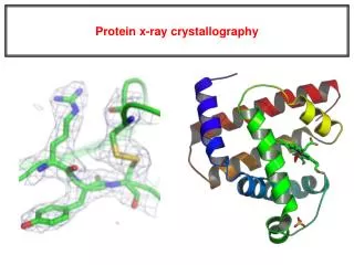
X-Ray Crystal Structures • Provide structural insights into protein mechanism at the atomic level • Mean resolution = 2 Angstrom (0.2 nm) • Membrane proteins are notoriously difficult to crystalize • Obtaining novel crystal structures is generally a long process that includes many strategies that do not work • No “one size fits all” strategy for crystallization (however GPCR structures are becoming more standardized) GPCR opsin, Park et al. 2008
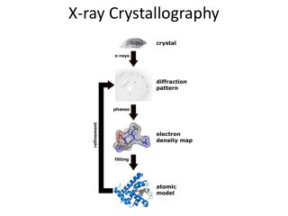
What is X-ray Crystallography? • Used to determine the arrangement of atoms within a crystal at high resolution • A beam of X-rays strikes the crystal and diffracts into many specific directions
What is X-ray Crystallography? • Based on the angles and intensities of the diffracted beam, a three dimensional structure of the density of electrons can be determined • From here, the mean positions and chemical bonds of the atoms within the crystal can be determined using mathematical Fourier transforms
Crystal Growth • With purified protein… • Cooling • Evaporation • Vapor Diffusion • Liquid Diffusion • Sublimation Crystals are typically larger than 0.1 mm
Membrane Proteins • Insertion of membrane proteins into the plasma membrane is driven by favorable helix-lipid interactions • Some polar transmembrane segments require help by neighboring proteins • Protein is not fully folded until it is inserted into the membrane • 75% of membrane proteins have a intracellular N-terminus • N-terminal tags aid in purification and do not hinder folding • Tags on extracellular N-terminus hinders translocation into membrane
Detergents for Membrane Proteins • During purification, detergents are used to solubilize membrane proteins • Contain polar head group and hydrophobic tail, just like phospholipid bilayer
Detergents for Membrane Proteins • During purification, detergents are used to solubilize membrane proteins Conventional Detergents, with polar head groups on right and hydrophobic chains at left Novel Detergents, amphiphiles contains “side” polar groups
Stabilizing Membrane Proteins • Point mutations • Example (Warne et al. and Serrano-Vega et al., 2008) • Alanine scanning mutagenesis used to stabilize the turkey B1-adrenergic receptor • Every residue was substituted with alanine and the effect of antagonist binding was measured at elevated temperatures to determine stabile mutant • Computational methods
Stabilizing Membrane Proteins • Membrane protein scaffolds • Fab fragments from monoclonal antibodies KvAP Crystal Structure stabilized by FAB fragments on the E2 Extracellular loop, Jiang et al 2003 FAB fragment KvAP
Types of Crystals • Type 1: 2D crystals with each membrane protein oriented side by sidewith hydrophobic surface of the protein initiating crystal contacts • Prepared in a lipid environment • Difficult to make • Type 2: Crystal contacts formed by the hydrophilic surface of membrane proteins, while the hydrophobic portions are shielded by detergent • Prepared in a detergent environment • Easier to make • The majority of membrane proteins have be determined with type 2 crystals.
Disadvantages for Membrane Proteins • Difficult to obtain stable membrane proteins in crystals without mutating the protein • Not in a native lipid bilayer, thus not a true native conformation • Crystal packing can include non-native conformation
Structural Variability at Neutral pH Neutral pH pH 4 (open)
Fluctuations in Crystal Structure Neutral pH pH 4 (open)
Changes in Functional Aspects of ECD Neutral pH pH 4 (open)
Overview Neutral pH pH 4 (open)
Kv1.2 • Voltage-dependent K+ channel • Member of the Shaker Kv Family • Contain highly conserved Pro-X-Pro signature sequence • Preceding the N-terminus, Shaker channels form a T1 domain • Beta2 subunit docks at the T1 domain • Contains an active site for NADP+ • Function not known, potentially regulates Kv activity
Kv1.2 Expression • Kv1.2 from rat was co-expressed with the B2 K channel beta subunit, in yeast Pichiapastoris • Channel contained a N-terminal 8x His tag and TEV protease cleavage site • N207Q mutation to elimination glycosylation at the S1-S2 linker • Preventing glycosylation may play reduce trafficking to membrane Pichia pastoris
To form Kv1.2 Crystals… • Protein was kept in a mixture of detergent and lipids • N-dodecyl-B-D-maltopyranoside or n-decyl-B-D-maltopyranoside) and 1-palmitoyl-2-oleoyl-sn-glycero-3-phosphocholine, 1-palmitoyl-2-oleoyl-sn-glycero-3-phosphoethanolamine and 1-palmitoyl-2-oleoyl-sn-glycero-3-[phospho-rac-(1-glycerol)] • Channel was kept in a strongly reducing environment using DTT and TCEP and in a oxygen depleted atmosphere
Crystals Formed via… • Vapor diffusion and frozen in liquid nitrogen
Kv1.2 Electron Density Electron Density Map = Blue VS = Voltage Sensor T1 = T1 Domain B = Beta Subunit T1 and Beta Subunit are more electron dense than pore and VS Mean B Factor T1 and B = 59 A2 Pore = 159 A2 VS = 162 A2 Based on electron density, a model of the channel was made
Kv1.2 Crystal Lattice Pore and VS (membrane spanning) = Red T1 and Beta Subunit (extramembranous regions) = Blue Due to low electron density, VS model built without connecting loops S1-S2, S2-S3 and S3-S4. S4-S5 was included
The Voltage Gated Potassium Channel Extracellular Intracellular
Kv1.2 Crystal Structure Pore VS S4-S5 linker 135 A NADP+ 95 A 95 A
Kv1.2 Crystal Structure Open Pore conformation VS 12 A Pore S4-S5 linker
Comparison of Kv1.2 to other K+ Channels Extracellular Intracellular Kv1.2 = Red (open pore) KcsA = Gray (closed pore) KvAP = Blue (open pore) * = Pro-Val-Pro
Charge Mapping of Kv1.2 Glutamate and aspartate residues = (-) charge = Red Arginine and lysine = (+) charge = Blue * = (-) charged Glu 128, Asp 129 and Glu 130 on T1 domain that interacts with positively charged amino acids on inactivation peptides for N-type inactivation (ball in chain)
Virtual Drug Screen • Exploit differences in GluN1 and GluN3 orthosteric binding site • Virtual screen of GluN3A LBD in antagonist bound state • 99 compounds obtained and functionally screened at GluN3A/B • Active hits evaluated at GluN1/2 • TK40
Antagonist Drug Potency Over Agonist 100 mM glutamate 0.5 mM glycine OR 100 mM glycine alone 3 mM glutamate 3000 mM glycine OR 10 mM glutamate alone