Download
1 / 21
210 likes | 364 Views
Immune system. Lecture 2011. Immune system. Innate - non-specific (no immunisation required) physical barriers (skin, mucosa, cilia) biological barriers (symbionts) chemical barriers (pH, mucus) soluble factors (lysozyme, interferons, proteins ac.ph., complement) Cells : phagocytes,

E N D
Immune system Lecture 2011
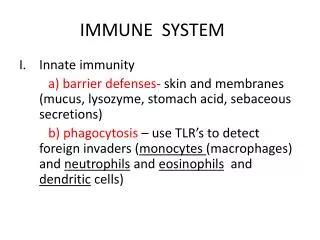
Immune system • Innate - non-specific (no immunisation required) • physical barriers (skin, mucosa, cilia) • biological barriers (symbionts) • chemical barriers (pH, mucus) • soluble factors (lysozyme, interferons, proteins ac.ph., complement) • Cells: phagocytes, granulocytes • (rapid answer, restrictive flexibility, non-specific reaction, no memory) • adaptive – specific (immunisation required) • Cells: T - lymfocytes (directly kill cells/ virus-infected, foreign cells, microorganisms) • B – lymfocytes (produce) • Antibodies (delayed answer, high flexibility, high specifity, memory and immunity)
Organs and cells of immune system Bone marrow Thymus Tonsils and adenoids Lymph nodes Spleen Peyer´s patches Appendix Lymphatic vessels internet
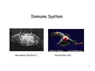
Cells of immune system (effect) • non-specific • intracelullar killingmacrophages (mononuclear phagocyte system) • “activating macrophages“! produce cytokins • APC! • neutrophils • extracellular killing • NK-cells (CD16, CD56), • “large, granular lymfocytes“ • (perforins, apoptosis), not MHC restricted • eosinophils (granules with cytotoxic proteins) • specific • B-lymphocytes (receptor: Ig) • T-lymphocytes (receptor:TCR in complex with CD, Ag split in peptide fragments in complex with MHC presented by APC (Tc)MHC I+Ag (TH)Ag +MHC II presenting by APC
Cell origin: Hemocytoblast (pluripotent stem cell) Myeloid lineage Erythrocytes Plateletes Granulocytes Monocytes Dendritic cells Mast cells Lymphoid lineage B-lymphocytes T-lymphocytes NK-cells
Tissues and organs of immune system • cells: blood, lymph, lymphoid tissue • lymphoid tissue: lymphoid nodules,MALT • primary or central lymphoid organs: thymus bone marrow • secondary or peripheral: encapsulated: lymph nodesspleen • non-capsulated: Peyer´s patches appendix tonsils
Cells of immune systemLYMPHOCYTES • Can exist without contact with another cells (cytokines!) • Migrate through tissues, blood and lymph • 2kg in organism/ 2-3 grams in blood
Lymphocytes NK
Cells of immune systemAntigen presenting cells (APC) • heterogenousgroup of cells macrophages dendritic cells Langerhans´ cells (skin) B-lymfocytes M-cells (GIT)
Dendritic cells • APC • originate in bone marrow, progenitor c. • precursors are seeded through the blood to (T-regions) or to non-lymphoid organs (Langerhans cc. in the skin) • high ability to be attracted to sites of antigen challenge and travel via lymph vessels to peripheral organs, presenting Ag to T-lymph (satelite lymph node, initiate immune response) • Xfolicular dendritic cc – origin just in stroma of nodes, not presenting Ag, but retain Ag/Ab in membrane – B-lymph and i. memory
Thymus • immature lymphocytes from bone marrow settled the thymus pre- and postnatally, undergoing -terminal differentiation and proliferation • elimination 95% (apoptosis), negative selection and positive selection • cortex (blood-thymus barries) x medulla (postcapillary venules – mature lymphocytes leave thymusto T-regions in peripheral organs) • reticular epithelial stroma, reticular cells! • Dual embryonic origin - endoderm (3rd pair of pharyngeal pouches) + mesenchym (lymphocytes), • Intensive growth till puberty • Inborn defect: di George syndrom- thymus aplasia
Thymus anatomy • Superior and anterior inferior mediastinum • lobus dx. et sin. • lobuli, cortex, medulla • (lobuli thymici accessorii) • weight at birth (12-14g)
Thymus – cortex (85% T-cells) • epithelial cells – cortical (stromal cells) • secretory granules,desmosomes,3D network, • express MHC I, MHC II • T-cells double negative, proliferation,gene rearrangement pre-TCR along with coreceptors CD4 and CD8 double positive (CD4 and CD8), positive selection( CD4 or CD8) • macrophages negative selection, apoptotic T-cc dendritic cells corticomedullary venules (functional thymocytes exit to circulation to T-regions
Thymus – medulla (25% T-cells) • Fully matured T-cells (single positive) • Epithelial cells • Hassal´s corpuscles (onion –like structures, degenerated cells • Macrophages • Dendritic cells • NO blood-thymus barrier
Blood – thymus barrier • Cortical epithelial cells • Basal lamina • Basal lamina • Endothelial cells • Macrophages • Only present in cortex
Thymus involution Gradual involution from puberty After 50th year, adipose tissue
Lymph node • organs of lymphoid tissue in the course of lymphatics • filter of Ag (microorganisms, tumor cells) coming in the lymph before its return to blood circulation • recirculation: lymphocytes return to node via high endothelial venules • reticular connective tissue stroma • cortex (lymphatic nodules, B-lymph) paracortex (T-lymph)
Lymph node Cortex: Subcapsullary sinuses Lymphatic follicules Interfollicular sinuses Paracortex Medulla Lymphatic cords Medullary sinuses
Spleen • largest lymphoid tissue accumulation • filter of Ag (microorganisms, tumor cells) that penetrate blood, producing antibodies and activated lymphocytes • White pulp , PALS (T-lymph) + lymhatic nodule (B-lymph) • Marginal zone (between red and white pulp, active macrophages) • Red pulp – lymphatic cords of Billroth + venous sinuses
Vascular supply Splenic artery Trabecular artery Central artery (surrounded by PALS) Penicilar artery (in red pulp) Venous sinuses Trabecular veins Splenic vein
Spleen – proliferation in germ center of lymhatic follicle (PCNA)