Download
1 / 84
940 likes | 1.58k Views
Chromosomes. Genetics 2005. Chromosomal Anomalies. Cytogenetics. The subdivision of genetics that focuses on chromosomes and cell division Abnormal cell divisions can lead to abnormal chromosomal numbers or other anomalies. Chromosome anomalies may cause phenotype abnormalities. .

E N D
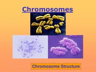
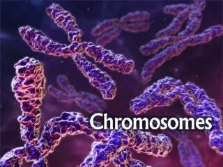
Chromosomes Genetics 2005
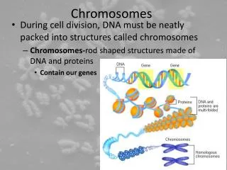
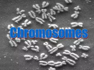
Cytogenetics • The subdivision of genetics that focuses on chromosomes and cell division • Abnormal cell divisions can lead to abnormal chromosomal numbers or other anomalies
Chromosome anomaliesmay cause phenotype abnormalities. • A chromosome karyotype revealed she carries three copies of chromosome 21, a condition called trisomy 21. Wendy Weisz has Down syndrome.
Karyotype • A chart arranging chromosome pictures according to their size, grouped by pairs of homologs. Chromosomes were named in order of their size and centromere position.
Anatomy of a chromosome • Centromeres are the largest constriction of the chromosome • Site of attachment of spindle fibers • 100,000s of 171 base pair repeat, called alpha satellite sequences • Centromere associated proteins are bound
Anatomy of a chromosome • Telomeres are: • At the tips of chromosomes • Many repeats of the sequence TTAGGG • Subtelomeres have more varied short repeats
Subtelomeres • The chromosome region next to the telomere consists of 8,000 - 300,000 bases including: • Repeats similar to the telomere sequence • Shorter repeats • Multigene families of genes • (e.g. olfactory receptor genes)
Anatomy of a chromosome • Chromosomes are categorized by the relative location of their centromere. • At tip - telocentric • Close to tip - acrocentric • At midpoint - metacentric • Displaced from center - • submetacentric
Largest, Metacentric Smallest acrocentric Sex chromosomes XX (shown)
Karyotype Metaphase chromosomes are squashed on a slide and stained with DNA binding dyes. Banding patterns help define different chromosomes.
Chromosome Map P arm Chromosome #1 Centromere Q arm
Chromosomal shorthand • An ideogram represents a chromosome schematically. • The major banding regions are indicated with numbers. • Sucrose intolerance is located at 3q.26 • (chromosome 3, long arm, major band 26)
FISH: fluorescence in situ hybridizationDNA probes labelled with fluorescing dye bind complementary DNA
telomeres centromeres
Visualizing chromosomes • Obtain tissue from person • Fetal tissue: amniocentesis • chorionic villi sampling • fetal cell sorting • Adult tissue: blood (white blood cells) • cheek swab (buccal cells) • skin cells • tissue biopsy • Prepare cells on slide to remove rest of cell matter • Stain DNA with dyes or DNA probes to visualize DNA • Evaluate chromosomes in comparison to known information
The normal karyotype: 23 diploid chromosomes • Human somatic cells contain 46 chromosomes: • paired homologs of chromosomes 1 to 22 and • sex chromosomes (XX or XY) • • Diploid refers to the presence of two copies of each different chromosome. • Gametes have one set of each chromosome and are called haploid. • Cells which contain a normal chromosome constitution are called euploid.
Polyploidy Aneuploidy monosomy trisomy Deletion Duplication Inversion Translocation Iso chromosome Ring chromosome Extra chromosome set Extra or missing chromosome one chromosome absent one chromosome extra Part of a chromosome missing Part of a chromosome present twice Segment of chromosome reversed Two chromosome arms exchanged in part or entirely A chromosome with identical arms A chromosome that forms a ring due to deletions in telomeres, which cause ends to adhere Chromosome Abnormalities
Polyploidy • Individuals with three copies of each chromosome are triploid. • Polyploidy accounts for 17% of all spontaneous abortions and 3% of stillbirths/newborn deaths. • Result of: • Two sperm fertilize one egg. • Haploid sperm fertilizes diploid egg.
Aneuploidy • Cells with extra or missing chromosomes are aneuploid. • Nondisjunction is a common cause of aneuploidy resulting in a gamete with one extra chromosome and another gamete with one missing chromosome. • Nondisjunction during the first meiotic division results in a copy of each homolog in the gamete. • Nondisjunction during the second meiotic division results in a both sister chromatids in one gamete.
Abnormal gametes Abnormal gametes Normal gametes Nondisjunction causes aneuploidy Nondisjunction in meiosis I Nondisjunction in meiosis I Anaphase I Nondisjunction in meiosis II Anaphase II Gametes
Anaphase I Homologs of small chromosome fail to separate Anaphase II Gametes Abnormal gametes Nondisjunction in meiosis I Sister chromatids separate normally monosomic trisomic
Anaphase I Homologs segregate normally Anaphase II Gametes Abnormal gametes Normal gametes Nondisjunction in meiosis II Sister chromatids fail to separate (left) trisomic monosomic
Turner syndrome • 45, X • 1 in 2,000 female births • 99% of Turner die in utero • Absence of Y leads to development as a female. • Absence of two copies of X-linked genes in a female results in Turner syndrome. • Phenotypes include short stature, webbing at back of neck, incomplete sexual development, hearing impairment.
Triplo-X aneuploidy • 47, XXX • 1 in 1,000 female births • Extra copy of every X-linked gene • Few modest effects on phenotype include tallness, menstrual irregularities and slight impact on intelligence • X-inactivation of two X chromosomes occurs while third remains active seems to compensate for presence of extra X.
Klinefelter syndrome • 47, XXY • 1 in 1,000 male births • Extra copy of each X-linked gene • Phenotypes include incomplete sexual development (rudimentary testes and prostate), long limbs, large hands and feet, some breast tissue development. • Some cases are not diagnosed until fertility problems arise or remain undiagnosed.
XYY syndrome • 47, XYY • 1 in 1,000 male births • Extra Y chromosome • 96% phenotypically normal • Modest phenotypes may include great height, acne and minor speech and reading problems. • Studies suggesting some increase in aggressive behaviors remain controversial.
Trisomies and Monosomies • One extra or one missing chromosome results in extra or missing copies of all of the genes on that chromosome. • Most trisomies and monosomies produce inviable embryos. • Some fetuses with trisomy of smaller autosomes survive to birth with syndromic conditions: