Download
1 / 63
930 likes | 2.19k Views
Introduction to the Assessment of Skeletal Dysplasias. M Skae & M Kaleem Bone & Calcium Disorders Annual Study Day 28 th Sept’12. Introduction and overview Assessment – before x-rays The basics of radiological interpretation Cardinal clues X-rays Who to involve – the MDT

E N D
Introduction to the Assessment of Skeletal Dysplasias M Skae & M Kaleem Bone & Calcium Disorders Annual Study Day 28th Sept’12
Introduction and overview • Assessment – before x-rays • The basics of radiological interpretation • Cardinal clues • X-rays • Who to involve – the MDT • Tools of the trade
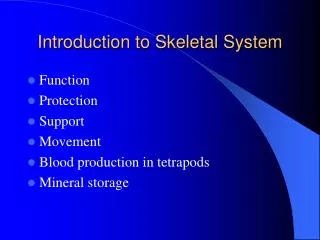
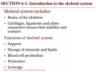
Introduction • Skeletal dysplasias are conditions with generalised skeletal abnormalities • Usually associated with disproportionate short stature, normal intelligence • Incidence 1/5000 live births • Classified on clinical, radiological and molecular criteria and sometimes histology
Overview • 2010 Nosology and Classification of Genetic Skeletal disorders • >450 different dysplasias, >220 genes • ~100 have prenatal onset • remainder presenting in infancy or age 2-3years • In some conditions, features disappear with time and therefore are more difficult to diagnose retrospectively in adults.
Radiological diagnostic groupings • Achondroplasia group • Metatropic dysplasia group • Short-rib polydactyly (SRP) group • Diastrophic dysplasia (DD) group • Type II Collagenopathies • SEMDs • Multiple epiphyseal dysplasia (MED) and pseudoachondroplasia group • Chondrodysplasia punctata (CDP) group • Metaphyseal chondrodysplasia (MCD) • Spondylometaphyseal dysplasia (SMD) group • Mesomelic dysplasia • Acromelic / acromesomelic group • Dysplasias with prominent membranous bone involvement – CCD • Bent bone dysplasia – Campomelic • Dysostosis multiplex group • Decreased bone density dysplasias – OI • Increased bone density dysplasias – osteopetrosis, pyknodysostosis • Defective mineralisation dysplasias – Hypophosphatasia • Craniotubular dysplasias – Pyle • Disorganised cartilagenous development – enchondrodysplasias • Osteolysis group • Patellar dysplasia – nail-patella syndrome
Normal Rhizomelic Mesomelic Micromelic Assessment I - disproportion Upper/lower segment ratio: • 1.7 newborn • 1.0 ages 2-8yrs • 0.95 adult Sitting height: ascertains trunkal shortening Limb lengths: • Rhizomelia (humerus and femur) • Mesomelia (radius, ulna, tibia and fibula) • Acromelia (Hands and feet) Body asymmetry Spine: assess for scoliosis, kyphosis and lordosis
Assessment II – General examination • General examination: facial features, hair quality, dental health, nails • Systemic features: renal problems, cardiac abnormalities • Developmental history: Most normal • Family history • Ethnicity: CHH in Amish, SEMD with joint laxity in SA • Joint pain
Assessment III - Radiology Skeletal survey: • Skull AP & Lateral • Spine AP & Lateral • Pelvis AP • 4 Limbs AP, occasional lateral Knee (assessment of patella) • Hands • Feet
Radiological assessment I • Epiphyseal dysplasia – small under ossified epiphyses • Metaphyseal dysplasia – widened, flared or irregular metaphyses • Diaphyseal dysplasia – cortical thickening or marrow space expansion or reduction
Radiological assessment II • Epiphyseal dysplasia • Metaphyseal dysplasia
Radiological assessment III • Vertebral (spondylo) abnormalities • Combinations: • Spondylo-epiphyseal dysplasia (SED) • Spondylo-metaphyseal dysplasia (SMD) • Metaphyseal-epiphyseal dysplasia (MED) • Spondylo-epiphyseal-metaphyseal dysplasia (SEMD)
Question - Is it acquired? Rule out acquired causes of bone problems: • Neuromuscular disorders • Chronic diseases – JIA • Poorly healed fractures • Metabolic bone problem
Question II – Is it a common dysplasia? Kozlowski and Beighton • Achondroplasia • Cleidocranial dysostosis • Dactyly - Brachydactyly , Camptodactyly , Polydactyly , Syndactyly • Enchondromatosis (Ollier) • Fibrous dysplasia • usual form (Jaffe-Lichtenstein) • with skin pigmentation and precocious puberty (McCune-Albright) • Gaucher's • Hypophosphatemic rickets • Marfan's • Multiple hereditary exostoses • Neurofibromatosis • Osteogenesis imperfecta • Osteopetrosis, pyknodysostosis • Osteopoikilosis INHERITANCE Autosomal dominant (but 50% new mutations) FGFR3 CLINICAL FEATURES Megalocephaly Short limbs Prominent forehead Thoracolumbar kyphosis Midfacial hypoplasia Short stature RADIOLOGY Diminishing interpeduncular distances between L1 and L5 COMPLICATIONS Short stature Dental malocclusion Hydrocephalus Repeated otitis media
Cardinal clues – cleidocranial dysostosis • Large head • Delayed suture closure • Hypertelorism, small face • Dental dysplasia – multiple teeth • Hypoplasia / aplasia of the clavicles
Cardinal clues – cartilage-hair hypoplasia • McKusick type metaphyseal chondrodysplasia • Short limbed dwarfism • Sparse hair • Autosomal recessive RMRP gene • T-cell and B-cell immunodeficiency • Dysplastic nails and brachydactyly • Notched incisors
Cardinal clues – Ellis van Creveld (chondroectodermal dysplasia) • Short stature, mesomelia • Narrow chest and short ribs • Polydactyly • Dysplastic nails • Dental abnormalities – missing teeth, lip fusion to gingiva • Cardiovascular abnormalities • AR – EVC1 & EVC2
Cardinal clues - Trichorhinophalyngeal syndrome (TRP) Type II / Langer-Giedion syndrome • Short stature • Unusual facies – long bulbous nose • Developmental delay • Cone epiphyses of the metacarpals • Bony exostoses especially distal tibia and ulna
Ear cysts • Hitchhiker thumb – shortened 1st metacarpal Diastrophic dysplasia • Pierre Robin sequence – midface hypoplasia, high arched palate, micrognathia • Myopia • Hearing problems Type II Collagenopathies
Cardinal clues - Osteopoikilosis • Dalmation disease – AD, LEMD3 & EXT1 • small round or oval foci of bone sclerosis located in the trabecular bone • particularly in the pelvis, metaphyses and epiphyses of long bones, tarsals, and carpals
Cardinal clues - Melorheostosis • Dripping wax appearance • LEMD3 mutations • Linked to osteopoikilosis • Buschke-Ollendorff syndrome – dermatofibrosis lenticularis
Cardinal clues - osteopetrosis • Extra dense bone • ‘Bone in bone’ appearance • Failure of normal osteoclast activity • May lead to marrow suppression – pancytopenia • Neural foramina stenosis
Cardinal clues - Enchondromatosis • Ollier’s syndrome • Not inherited • central expansile pattern or linear metaphyseal lucencies • 5-30% malignant degeneration to chondrosarcoma • higher risk if associated with soft tissue haemangiomas (Mafucci's syndrome)
Who to involve - The MDT • Geneticist • Radiographer • Metabolic bone doctor • Orthopaedic surgeon • Spinal surgeon • Physiotherapist • Occupational therapist
Tools of the trade • A good atlas – Spranger, Brill and Poznanski • Warman et al. Nosology and classification of genetic skeletal disorders: 2010 revision. American Journal of Medical Genetics 2011 May;155(5): 943–968, May 2011 • Alanay & Lachman. A Review of the Principles of Radiological Assessment of Skeletal Dysplasias • J Clin Res Pediatr Endocrinol. 2011 December; 3(4): 163–178 • Unger et al. A diagnostic approach to skeletal dysplasias. Paediatric Bone Disease 2003, 16. Elselvier Science.
Phone or e-mail a friend! • The European Skeletal Dysplasia network (ESDN) – usually accessed by the radiologists or genetists • The North-western Skeletal Dysplasia Group
Musa Kaleem (MBBS, MRCPCH, FRCR) Imaging Skeletal Dysplasias
Constitutional disorders of bone osteochondrodysplasias dysostoses • Dysplasias (growth) • Osteodystrophies (texture) • Failure of gene expression • Phenotype usually continues to evolve • Defective bone formation due to a defect in blastogenesis • Remain static • do not spread to involve normal bones Offiah et al; Pediatr Radiol 2003
Zones • Resting • Proliferating cartilage • Hypertrophic cartilage • Provisional calcification • Ossification
Genetics Skeletal Survey • Skull (AP & Lat) • Spine (AP & Lat) • Chest • Pelvis • One upper limb • One lower limb • Left hand (bone age) • Additional views • Lateral knee for assessment of patella) • Lateral foot (for calcaneum) • Foetogram/ babygram • AP • Lateral
Radiological assessment – stepwise approach • Step 1 – assessment of disproportion • Spine • limb segments (rhizo/ meso/ acro) • Step 2 – assessment of epiphyses, metaphyses and diaphyses • Step 3 – assessment of bone density / texture
Radiological assessment (2) • Step 4 – search for other clues • Skull • Cranio-cervical junction • Spine • Ribs/ clavicles • Pelvis • Long bones • Hands and feet • Step 5 – Seek help from colleagues/ refer to textbook/ Electronic database
Radiological diagnostic groupings • Achondroplasia group • Metatropic dysplasia group • Short-rib polydactyly (SRP) group • Diastrophic dysplasia (DD) group • Type II Collagenopathies • SEMD • Multiple epiphyseal dysplasia (MED)and pseudoachondroplasia group • Chondrodysplasiapunctata (CDP) group • Metaphysealchondrodysplasia (MCD) • Spondylometaphyseal dysplasia (SMD) group • Mesomelic dysplasia • Acromelic / acromesomelic group • Dysplasias with prominent membranous bone involvement – CCD • Bent bone dysplasia – Campomelic • Dysostosis multiplex group • Decreased bone density dysplasias – OI • Increased bone density dysplasias – osteopetrosis, pyknodysostosis • Defective mineralisation dysplasias – Hypophosphatasia • Craniotubulardysplasias – Pyle • Disorganised cartilagenous development – enchondrodysplasias • Osteolysis group • Patellar dysplasia – nail-patella syndrome Alanay Y, Lachman RS et al; J Clin Res Pediatr Endocrinol: 2011
Radiological hints to diagnoses Skull • Changes in density, size and shape • Wormian bones • OI • Cleidocranialdysostosis • Pyknodysostosis • Craniosynostosis • Crouzon’s/ Pfeiffer’s • Skull base/ midface hypoplasia • Basilar invagination
Generalised reduced density: Osteogenesis Imperfecta (OI) Glass R B J et al. Radiographics 2004;24:507-522
Wormian bones… They are named after Ole Worm, a Danish anatomist who described them From radiopedia.org
Increased density: osteopetrosis • AR • Benign vs malignant forms • Presents with infection/ cranial nerve palsies
Increased density: generalised Pycnodysostosis
Frontometaphyseal dysplasia Frontometaphyseal dysplasia Glass R B J et al. Radiographics 2004;24:507-522
Spine • Odontoid hypoplasia/ atlanto-axial subluxation • Kyphoscoliosis (gibbus) • Pedicles (length/ interpediculate distance) • Vertebral body shape abnormalities • Platyspondyly • Bullet shaped vertebrae/ vertebral beaking • Scalloped vertebrae • Humps • Cleft vertebrae (sagittal/ coronal)
CDPX2 Chondrodysplasia Punctata (x-linked)