Download
1 / 70
700 likes | 736 Views
Learn about the four types of tissues: epithelial (types and locations, functions, classifications) and connective (functions, types, characteristics).

E N D
Principal Types of Tissue Four types of tissues: I. Epithelial tissue II. Connective tissue III. Muscle tissue IV. Nervous tissue
Extracellular Matrix (ECM) • Functions • Helps bind tissues together structurally • Allows local communication among ECM and various cells—through connection via integrins in plasma membranes
Extracellular Matrix (ECM) • Components • Water • Proteins • Structural proteins • Collagen—strong, flexible protein fiber • Elastin—elastic fibers • Includes glycoproteins—proteins with a few carbohydrate attachments • Glycoprotein attachments also allow local communication within a tissue
I. Epithelial Tissue • Functions • Protection • Sensory functions • Secretion • Absorption • Excretion
I. Epithelial Tissue • Types and locations • Epithelium is divided into two types: • Membranous epithelium—covers the body and some of its parts; lines the serous cavities, blood and lymphatic vessels, and respiratory, digestive, and genitourinary tracts • Glandular epithelium—secretory units of endocrine and exocrine glands
I. Epithelial Tissue • Classification of epithelial tissue • Classification based on cell shape • Squamous • Cuboidal • Columnar • Pseudostratified columnar
I. Simple epithelium 1.Simple squamous epithelium • One-cell layer of flat cells • Permeable to many substances • Examples: endothelium—lines blood vessels; mesothelium—pleura
I. Simple epithelium 2. Simple cuboidal epithelium • One-cell layer of cuboidal cells • Found in many glands and ducts
I. Simple epithelium 3. Simple columnar epithelium • Single layer of tall, column-shaped cells • Cells often modified for specialized functions—e.g., goblet cells (secretion), cilia (movement), microvilli (absorption) • Often lines hollow visceral structures
I. Simple epithelium 4. Pseudostratified columnar epithelium • Columnar cells of differing heights • All cells rest on basement membrane but may not reach the free surface above • Cell nuclei at odd and irregular levels • Found lining air passages and segments of male reproductive system • Motile cilia and mucus are important modifications
I. Stratified epithelium 5. A. Stratified squamous (keratinized) epithelium • Multiple layers of flat, squamous cells • Cells filled with keratin • Covers outer skin on body surface
I. Stratified epithelium 5. B. Stratified squamous (nonkeratinized) epithelium • Lines vagina, mouth, and esophagus • Free surface is moist • Primary function is protection
I. Stratified epithelium 6. Stratified cuboidal epithelium • Two or more rows of cells are typical • Basement membrane is indistinct • Located in sweat gland ducts and pharynx
I. Stratified epithelium 7. Stratified columnar epithelium • Multiple layers of columnar cells • Only most superficial cells are typical in shape • Rare • Located in segments of male urethra and near anus
I. Stratified epithelium 8. Stratified transitional epithelium • Located in lining of hollow viscera subjected to stress (e.g., urinary bladder) • Often 10 or more layers thick • Protects organ walls from tearing
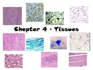
Can you identify these? B A E C D F G
II. Connective Tissue • Functions, characteristics, and types • General function—connects, supports, transports, and protects • General characteristics—extracellular matrix (ECM) predominates in most connective tissues and determines its physical characteristics; consists of fluid, gel, or solid matrix, with or without extracellular fibers (collagenous, reticular, and elastic) and proteoglycans or other compounds that thicken and hold together the tissue
II. Connective Tissue • Four main types: 1.Fibrous A. Loose, ordinary (areolar) B. Adipose C. Reticular D. Dense • Irregular • Regular (collagenous and elastic) • 2. Bone • A. Compact bone • B. Cancellous bone • 3. Cartilage • A. Hyaline • B. Fibrocartilage • C. Elastic • 4. Blood
1. Fibrous connective tissue A. Loose, ordinary (areolar) connective tissue • One of the most widely distributed of all tissues • Intercellular substance is prominent and consists of collagenous and elastic fibers loosely interwoven and embedded in soft, viscous ground substance Function—stretchy, flexible connection
1. Fibrous connective tissue B. Adipose tissue • Similar to loose connective tissue but contains mainly fat cells • Functions—protection, insulation, support, and food reserve
1. Fibrous connective tissue C. Reticular tissue • Forms framework of spleen, lymph nodes, and bone marrow • Consists of network of branching reticular fibers with reticular cells overlying them • Functions—defense against microorganisms and other injurious substances; reticular meshwork filters out injurious particles, and reticular cells phagocytose them
1. Fibrous connective tissue D. Dense fibrous tissue • Matrix consists mainly of fibers packed densely and relatively few fibroblast cells • Irregular—fibers intertwine irregularly to form a thick mat (Figure 5-20) • Regular—bundles of fibers are arranged in regular, parallel rows • Collagenous—mostly collagenous fibers in ECM (Figure 5-21 and 5-22) • Elastic—mostly elastic fibers in ECM (Figure 5-23) • Locations—composes structures that need great tensile strength, such as tendons and ligaments; also dermis and outer capsule of kidney and spleen • Function—furnishes flexible connections that are strong or stretchy
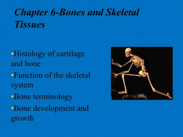
2. Bone tissue • Highly specialized connective tissue type • Cells—osteocytes—embedded in a calcified matrix • Inorganic component of matrix accounts for 65% of total bone tissue • Functions: • Support • Protection • Point of attachment for muscles • Reservoir for minerals • Supports blood-forming tissue
A. Compact bone • Osteon (Haversian system) • Structural unity of bone • Spaces for osteocytes called lacunae • Matrix present in concentric rings called lamellae • Canaliculi are canals that join lacunae with the central Haversian canal • Cell types: • Osteocyte—mature, inactive bone cell • Osteoblast—active, bone-forming cell • Osteoclast—bone-destroying cell • Formation (ossification) • In membranes—e.g., flat bones of skull • From cartilage (endochondral)—e.g., long bones, such as the humerus
B. Cancellous bone • Trabeculae—thin beams of bone • Supports red bone marrow • Myeloid tissue—a type of reticular tissue • Produces blood cells • Called spongy bone because of its spongelike appearance
3. Cartilage • Chondrocyte is only cell type present • Lacunae house cells, as in bone • Avascular—therefore, nutrition of cells depends on diffusion of nutrients through matrix • Heals slowly after injury because of slow nutrient transfer to the cells • Perichondrium is membrane that surrounds cartilage
3. Types of Cartilage A. Hyaline • Appearance is shiny and translucent • Most prevalent type of cartilage • Located on the ends of articulating bones B. Fibrocartilage • Strongest and most durable type of cartilage • Matrix is semirigid and filled with strong, white fibers • Found in intervertebral disks and pubic symphysis • Serves as shock-absorbing material between bones at the knee (menisci) C. Elastic • Contains many fine, elastic fibers • Provides strength and flexibility • Located in external ear and larynx
4. Blood • A liquid tissue • Contains neither ground substance nor fibers • Composition of whole blood • Liquid fraction (plasma) is the matrix—55% of total blood volume • Formed elements contribute 45% of total blood volume • Red blood cells, erythrocytes • White blood cells, leukocytes • Platelets, thrombocytes
4. Blood (cont.) • Functions • Transportation • Regulation of body temperature • Regulation of body pH • White blood cells destroy bacteria • Circulating blood tissue is formed in the red bone marrow by a process called hematopoiesis; the blood-forming tissue is sometimes called hematopoietic tissue
III. Muscle Tissue • Types (Table 5-7) 1. Skeletal, or striated voluntary (Figure 5-32) 2. Smooth, or nonstriated involuntary, or visceral (Figures 5-33 and 5-34) 3.Cardiac, or striated involuntary (Figure 5-35)