Download

1 / 38
380 likes | 403 Views
Learn about the different types of tissues in the body, including epithelial and connective tissues. Explore their functions, characteristics, and where they are found in the body. Dive into histology and the vital roles tissues play in the organ systems of the human body.

E N D
Remember…… Chemical Cellular Tissue Organ Organ System Organismal Level
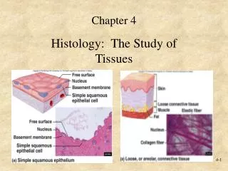
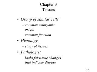
Tissues are: • groups of cells that are similar in structure and function • Histology = study of tissues • The four tissue types are: • Epithelial • Connective • Muscular • Nervous
Epithelial Tissue • Functions • protection: covers surfaces • sensory input • absorption • filtration • secretion
Epithelial Tissue • Characteristics • avascular • fit closely together • has a “top” and “bottom”: • apical surface (free) • basement membrane • regeneration
Epithelial Tissue: Classification • Number of cell layers • Simple • Stratified • Pseudostratified • Shape of apical surface cells • Squamous • Cuboidal • Columnar • Transitional
Epithelial Tissue: Simple Squamous Function: Good for Diffusion Where found? Figure 4.3
Epithelial Tissue: Simple Columnar Function: absorption and secretion Where found? Figure 4.5a
Epithelial Tissue: Pseudostratified Ciliated Columnar Function: secrete mucus and move mucus with cilia Where found?
Epithelial Tissue: Stratified Squamous Function: protection from abrasion Where found?
Epithelial Tissue: Transitional Functions: stretches Where found?
Epithelial Tissue: Simple Cuboidal Functions: secretion Where found? Figure 4.4a
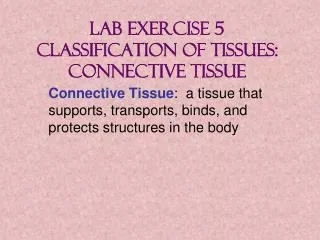
Connective Tissue • Functions • protection • support • bind together other tissues of body • Most abundant and widespread tissue in body
Connective tissue • Characteristics • Rich blood flow • Exceptions? • Composed of • Specialized cells and • Extracellular Matrix (nonliving area between cells) • protein fibers & ground substance
Four Connective Tissue Types 2 3 4 1 Fibrous Liquid Hard
Connective Tissue: Areolar Cells: fibroblasts Matrix: gel-like Function: wraps and cushions organs
Connective Tissue: Adipose Cells: Adipocytes (fat) Matrix: sparse Function: reserves, protection, insulation Reticular Tissue Figure 4.11
Connective Tissue: Dense Regular Cells: fibroblasts Matrix: primarily collagen fibers Function: tendons and ligaments
Connective Tissue: Hyaline Cartilage Cells: chrondrocytes in lacunae (egg-shaped) Matrix: firm with collagen Function: support and reinforce (ribs, nose, trachea)
Elastic Cartilage • More flexible/pliable • Supports and protects outside the bone • Where found?
Fibrocartilage • More dense than other cartilages • Good for support, cushioning, and shock absorption between bones • Pubic Symphysis, Meniscus of knee, verterbral column
Connective Tissue: Bone Cells: osteocytes in lacunae Matrix: hard, calcified with collagen Function: support and attachment
Blood & Lymph Fluid • WHY is this liquid tissue considered a connective tissue? • What is its function?
Muscle Tissue • 3 Types of Muscle Tissue • Skeletal • Smooth • Cardiac • Function of each?
Nerve Tissue • Function?
Integumentary System • Skin • Epidermis • Epithelial Tissue (Stratified squamous) • Dermis • Connective Tissue • Hypodermis (superficial fascia) • Connective Tissue
Hair shaft Pore Dermal papillae (papillary layer of dermis) Epidermis Meissner's corpuscle Free nerve ending Reticular layer of dermis Sebaceous (oil) gland Arrector pili muscle Dermis Sensory nerve fiber Eccrine sweat gland Pacinian corpuscle Artery Hypodermis (superficial fascia) Vein Adipose tissue Hair root Hair follicle Eccrine sweat gland Hair follicle receptor (root hair plexus) Figure 5.1
Epidermis Which layer is missing? 4 Cell Types: Keratinocytes Langerhans cells Melanocytes Merkel cells
The Structure of the Epidermis Figure 5.4
Skin (Integument) Figure 5.1
Structure of the Dermis Meissner’s corpuscles Pacinian corpuscles
Accessory Organs of the Skin • Hair • Nails • Sebaceous Glands • Sudoriferous (Sweat) Glands
Sebaceous Glands and Follicles • Produce sebum (oil) • Ducts empty into hair follicle Figure 5.11
Sweat (Sudoriferous) Glands Apocrine: found in axillary & anogenital areas Figure 5.12a, b
Sweat (Sudoriferous) Glands Eccrine (Merocrine) • found in palms, soles of the feet, and forehead • water, salt and urea Figure 5.12a, b


![[Insert exercise name]](https://cdn0.slideserve.com/1400721/insert-exercise-name-dt.jpg)