Download
1 / 30
310 likes | 566 Views
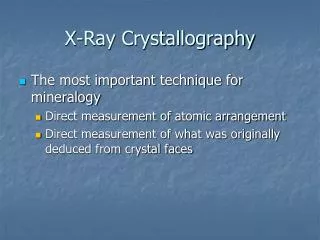
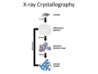
X-ray Crystallography. GLY 4200 Fall, 2014. Discovery of X-rays. Wilhelm Conrad Roentgen discovered x-radiation in 1895 In 1912, Friedrich, Knipping, and von Laue demonstrated diffraction of x-radiation passing through a crystal The wavelength of x-radiation ranges from 10 -6 to 10 -1 nm.

E N D
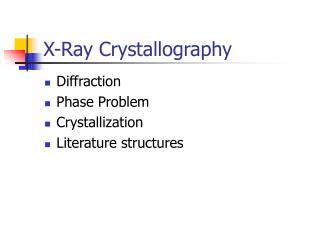
X-ray Crystallography GLY 4200 Fall, 2014
Discovery of X-rays • Wilhelm Conrad Roentgen discovered x-radiation in 1895 • In 1912, Friedrich, Knipping, and von Laue demonstrated diffraction of x-radiation passing through a crystal • The wavelength of x-radiation ranges from 10-6 to 10-1 nm
Einstein Equation • E = hυ = hc/λ • where • E = energy • h = Planck's constant • υ = frequency • c = speed of light • λ = wavelength.
Conversion to Kinetic Energy • If all the kinetic energy of an electron is converted to X-ray quanta, we can rewrite the equation as: • eV = hc/λ • Replacing constants gives: • λ(nm) = 1.24/kV • Where kV = kilovolts
White Radiation • Effect of excitation potential on minimum wavelength
X-ray Tube • X-ray tube schematic diagram
Electron Shells • Electron infall from outer to inner shells
Copper Energy Levels • Energy-level diagram for electron transitions in Cu
Absorption Edge • Absorption edge of Ni in relation to the emission spectrum of Cu
Scattering • Scattering of X-rays by a row of equally spaced, identical atoms
Reflection • Condition for reflection
Path Difference • Path difference = 2d sin θ
Bragg Equation • nλ = 2d sin θ • where • n is an integer • d is the distance between successive parallel planes (the "interplanar" spacing) • θ = glancing angle of incidence • This is the condition for successful reinforcement of waves reflected off different layers
W.H. and W.L. Bragg • Derived by English physicists Sir William Henry Bragg and his son Sir William Lawrence Bragg • Shared Nobel Prize in Physics, 1915
Diffracted X-ray Cones • Diffraction cones from a row of atoms
Cone Intersection • Diffraction cones from three noncoplanar rows of scattering atoms, intersecting in a common line
Figure 12 • Arrangement for a powder photograph
Powder Pattern • Diagram showing the formation of lines from a powder
Laue Method • a) Obtaining a Laue photograph with a stationary crystal • b) Laue photograph of vesuvianite, taken along the A4 axis. Axial directions a1 and a2 were inked onto the photograph after development.
Laue Film • Laue photograph, mineral unknown • Named for its developer German physicist Max von Laue, who won the Nobel Prize in Physics in 1914 for the discovery of diffraction of X-rays by crystals
Weissenberg Rotation Method • Austrian physicist Karl Weissenberg developed a rotating-crystal method which also translated the film, allowing unambigious index of each refraction
Precession Camera • Buerger precession camera
Martin Julian Buerger • American Crystallographer who developed the precession camera • Crystal and the film move • Film shows an undistorted replica of the corresponding reciprocal lattice plane • Each diffraction may be indexed
Precession Film of Wavellite • A precession photograph is quickly indexed since it shows very clearly the symmetry content of the reciprocal lattice • Indeed the distance between the spots on the film is simply the reciprocal lattice distance between two nodes, scaled by the X-ray wavelength and the camera radius
Four Circle Diffractometer • A crystal is randomly set on the goniometric mount • Computer will measure and calculate the exact value that each of four angles has in order to observe the reflections of a specific set of planes (hkl)
Mounting Methods for Powders • Placed in fine capillary tube of 0.2mm bore • Coated on a fine glass fiber - the fiber is dipped in a liquid such as alcohol and then rolled in the powder • Mixed with gum arabic and rolled between slips of glass into a fine spindle or a tiny ball, no more then 0.3 mm in diameter • Sprinkled on a piece of tape mounted over a hole drilled in a circular piece of metal
Powder Method Advantages • Fast - an analysis is completed in two hours or less • Requires very small sample amounts, which is especially important in cases where the material is rare • Sample preparation times are usually small • The cost per analysis is low, although there is an initial investment in the X-ray equipment and associated computer
Exam Date and Time • Lecture Final Examination • Friday, December 5, 2014 from 7:45 a.m. to 10:15 a.m.