Download
1 / 51
540 likes | 756 Views
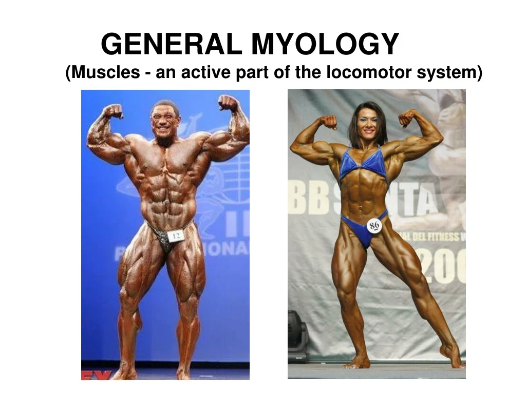
GENERAL MYOLOGY (Muscles - an active part of the locomotor system ). General function of muscles Produce movement in sites of skeletal junctions C hange shapes of body cavities and openings G ive information about the body position in 3D space

E N D
GENERAL MYOLOGY (Muscles - an active part of the locomotor system)
General function of muscles • Produce movement in sites of skeletal junctions • Change shapes of body cavities and openings • Give information about the body position in 3D space • Important role during thermoregulation • Help toblood and lymphcirculation • Verbal and non verbal comunication • Logistic system (supports respiration, digestion…) About 600 muscles (♂ 35%, ♀ 32% of weigth) Smooth musculature forms walls of vessels and hollow organs, works without our will, without fatigue Srdeční svalovina tvoří srdce, pracuje bez závislosti na naší vůli, po celý život, bez únavy Svalovina příčně pruhovaná, tvoří kosterní svalstvo, pracuje v závislosti na naší vůli, snadno se unaví, spotřebuje hodně energie, při práci tvoří teplo
On thebasisofstructure and physiologicalcharacteristicswedistinguish: 1) Striated(skeleton) muscles– musculisceleti(skeletalmuscles) + skin muscles(musculicutanei) (formmusclesoflimbs, workundercontrolofourwill, easyfatigued, spend a lot ofenergy, produce a heat) 2) Cardiacmuscles (myocardium) 3) Non-striatedvisceral (smooth) muscles–form an integralpart of some hollow organs and cavities, workwithoutourwill, withoutfatigue.
The main of the mechanical function of muscle fibers is shortening - contraction (movement). myocytes myofibrilscontractile proteins myosin and actin
Common structure of muscle Origo (origin) Fascia (cover) Tendo, aponeurosis Insertio (insertion) Caput (head) Venter(belly) Cauda (tail)
Structure of muscle Fascia –a fibrous membrane –separates the muscles (or groups) from adjacent structures. Vessels and nerves enter into muscle by its hilus(rich ramification) ORIGO fascia tendo INSERTIO (insertion) Tendons are attached to the bones by Sharpey´s fibres.
Innervation of muscles Cranial nerves(III. – XII.) Dorsal branches Spinal nerves (31 pairs) Ventral branches (form plexus) Cervical plexus Brachial plexus Intercostal nerves Lumbal plexus Sacral plexus
Auxiliary facilities 1. Fasciae– allow to move one muscle (or functional group) against the other; septa 2. Bursae synoviales (synovial bursae)– protect tendons against friction 3.Tendo aponeurosis=tendon of flat muscles 4.Trochleae musculares (muscular trochleae)– fibrous loops keeping tendon to a bone, permit change of direction of muscle pulling 5. Ossa sesamoidea (sesamoid bones)– at the places of pressure 6. Vaginae tendinum(tendon sheats)
Vaginae tendinum andvaginae synoviales (tendon´s and synovial sheaths) A space along tendons, closed, increasing sliding capacity of tendons stratum fibrosum =fibrous layer (Osteofibrous canal) stratum synoviale= (synovial layer) ext. and int. layerwith mesotenonium for penetration of vessels into tendon) Sharpey´s fibers Purulent inflammation can spread here
Division of muscles according to their shape • long type (predominantly limbmuscles) • short type (circumarticular muscles, shortmuscles • ofthe hand.....) • flat type (for eg. abdominal wallmuscles) • Composed: • muscleswith two or more heads: biceps, triceps, • quadriceps • multi-belliedmuscles: musculus digastricus(digastric muscle) • various types of sphinctersordilatators: mm. orbiculares(orbicular muscles), m. dilatatorpupilae • unipennate muscles or multipennate muscles:
Division of muscles according to the function synergists x antagonists flexors x extensors Example: biceps of brachium x triceps of brachium abductors x adductors Example: abductor pollicis brevis x adductor pollicis dilatators x sfincters Example: dilatator pupillae x sphincter pupillae
SPECIAL MYOLOGY Description of the muscle: • Name of muscle • Group (a part of body) Origo - origin Insertio - insertion Functio – function/action Innervatio - innervation
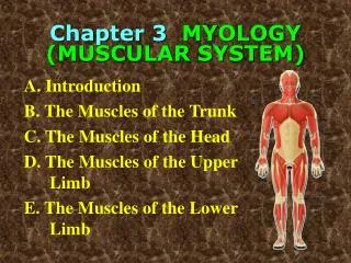
Division of muscles according to regions of the body Muscles of the head Muscles of the neck Muscles of the thorax Muscles of the abdomen Muscles of the diaphragma pelvis Muscles of the back Muscles of the upper limb Muscles of the lower limb
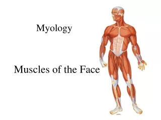
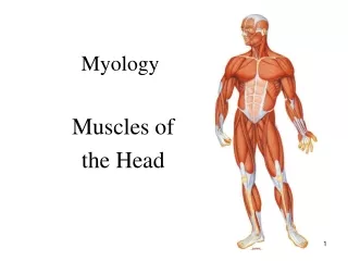
SPECIAL MYOLOGY Mm. capitis (Muscles of the head) • MUSCULI MASTICATORII (MASTICATORY MUSCLES) Innervation - n. trigeminus = V. cranial nerve 2) MUSCULI FACIALES (MUSCLES of FACIAL EXPRESSION) Innervation - n. facialis = VII. cranial nerve NO FASCIA!– skin muscles
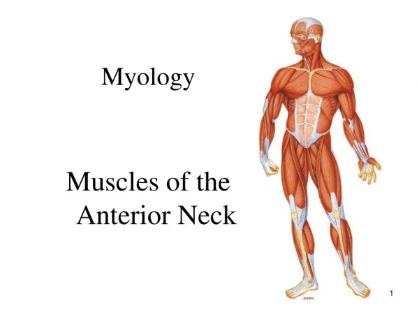
Musculi colli – muscles of the neck Superficial layer m. platysma m. sternocleidomastoideus mm. suprahyoidei(depression of mandible) mm. infrahyoidei - mainly fixation of os hyoideum (hyoid bone) Deep layer - mainly flexion of the neck (and head) mm. scaleni mm. prae- and intervertebrales
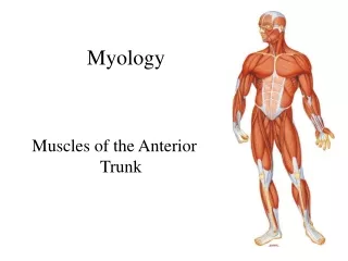
Musculithoracis, abdominis et dorsi (Musclesofthechest, abdomen and back)
Musculithoracis(thoracicmuscles) I. Thoracohumeralmuscles (mainlyventralflexion and abductionoftheupper limb Musculus pectoralis major (greater pectoral muscle) Musculus pectoralis minor (lesser pectoral muscle) Musculus subclavius Musculus serratus anterior II. True (intrinsic) thoracic muscles muscles for respiratory movements Musculi intercostales externi, interni et intimi Musculus transversus thoracis III. Diaphragma mainmuscle for inspiration
II. True (original=intrinsic) thoracic muscles muscles for respiratory movements 1. Mm. intercostales externi 2. Mm. intercostales interni 3. Mm. intercostales intimi 4. M. transversus thoracis
Musculiabdominis (abdominalmuscles) antagonistsofthedorsalmuscles, regulatethevolumeoftheabdominalcavity
Musculiabdominis(musclesofthe abdomen) antagonistsofthedorsalmuscles, regulatethevolumeoftheabdominalcavity Ventral group musculus rectus abdomis (+ its sheat=vagina mm. recti abdominis) musculus pyramidalis Lateral group musculus obliquus externus abdominis musculus obliquus internus abdominis musculus transversus abdominis musculus cremaster Dorsal group musculus quadratus lumborum
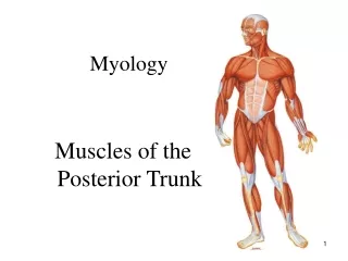
Musculi dorsi (muscles of the back) I. Extrinsicmusclesoftheback II. Intrinsicmusclesoftheback(locateddeeper, innervation by dorsalramiofspinalnerves) III. Shortmusclesoftheback Ad I. Extrinsicmusclesoftheback A) Mm. spinohumerales(spinohumeralgroup)movementsoftheupper limb B) Mm. spinocostales(spinocostalgroup)helprespiratorymovements
I. EXTRINSIC BACK MUSCLES A. Mm. spinohumerales (spinohumeral group) 1. m. trapezius 2. m. latissimus dorsi 3. m. levator scapulae 4. m. rhomboideus minor 5. m. rhomboideus major
B) Spinocostal group of muscles help respiratory movements 1. m. serratus posterior superior 2. m. serratus posterior inferior
Ad II. Intrinsicmusclesoftheback mainlyextensorsoftheback and thehead, innervationramidorsalesofspinalnerves 1)Spinotransversalsystem(m. spleniuscapitis and cervicis) 2) Sacrospinalsystem(m. erectorspinae, longissimus and iliocostalis) 3) Spinospinalsystem(m. spinalisthoracis) 4) Transversospinalsystem(m. semispinaliscapitis and cervicis)
Ad III. Short muscles of the back Mm. nuchae profundi a)m.rectus capitis posterior minor(lesser) b) m. rectus capitis posterior major (greater) c) m. obliquus capitis superior d) m. obliquus capitis inferior
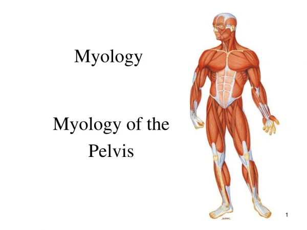
Diaphragma pelvis m. levator ani (m. pubococcygeus, m. iliococcygeus) m. coccygeus Lifting and closure of rectum Diaphragma urogenitale (ventrally + caudally) m. transversus perinei profundus (+ m. sphincter urethrae) m. transverus perinei superficialis Muscles of the external sexual organs: m. ischiocavernosus m. bulbospongiosus m. sphincter ani externus
Musculi masticatorii (innervation by n. trigeminus = V. cranial nerve) • M. temporalis(temporalmuscle) • M. masseter(massetermuscle) 3. M. pterygoideusmedialis (medialpterygoidmuscle) 4. M. pterygoideuslateralis (lateralpterygoidmuscle)
Musculus temporalis (temporal muscle) Origin:linea temporalis inferior, fossa temporalis, fascia temporalis Insertion: processus coronoideus mandibulae (coronoid proces of mandible) Function:elevation of mandible, retraction Inervation:trigeminal nerve (n. V.)
Musculus masseter(masseter muscle) Origin:corpus ossis zygomatici, arcus zygomaticus (body and zygomatic arch) Insertion:tuberositas masseterica (masseteric tuberosity) of mandible Function:elevation and protraction of mandible Innervation:trigeminal nerve (n. V.)
Musculus pterygoideus medialis(medial pterygoid muscle) Origin:fossa pterygoidea and tuber maxillae Insertion:tuberositas pterygoidea Function:elevation of mandible and movements to the sides Innervation:trigeminal nerve (n.V.)
Musculus pterygoideus lateralis(lateral pterygoid muscle) Origin:facies infratemporalis of ala major ossis sphenoidalis and lamina lateralis processus pterygoidei Insertion:fovea pterygoidea mandibulae + discus and articular capsule of temporomandibular joint Function:protraction of mandible, chewing movements Innervation: trigeminal nerve (n.V.)
Muscles of mastication M. masseter and m. pterygoideus medialis form a „loop“ around mandible
Mm. faciales(muscles of facial expression) facial nerve – n.VII. Muscles of the scalp Muscles of the orbit region Muscles of the nasal region Muscles of the mouth region Their contraction causes shift of the skin (folds or wrinkles) – it is the basis of the facial expression. They have no fascias!
Muscles of the scalp M. Epicranius M. occipitofrontalis with venter frontalis and occipitalis (frontal and occipital belly) o: venter occipitalis from highest nuchal line (linea nuchae suprema) i: galea aponeurotica o: venterfrontalis from galea aponeurotica i: skin of eyebrow and glabellar region f: wrinkles in the forehead, lift the eyebrow (astonishment) M. temporoparietalis Rudimentary muscle, from galea aponeurotica to auricular cartilage
Muscles of the region of the palpebral fissure M. orbicularis oculi: Palpebral part, orbital part, lacrimal part Circularly around the orbit, basis of the eye lids, a sphincter. O: palpebral lig., frontal process of maxilla and lacrimal crests F: firmly closure the eyelid, palpebral part concerned with blink reflex, radial folds in lateral angle (expression of worry and concern) Corrugator supercilii: O: From the glabellar and supraorbital region I: skin of eyebrows F: produces a vertical wrinkles Procerus O: dorsum nasi (dorsum of nose) I: skin of the forehead F: transverse folds across the root of nose Nasalis O: alveolar juga of the canine tooth and lateral incisor I: skin of the side of the nose F: pulls the nasal wings downward and backward and reduce size of nostrils. Gives impression of desiring, demanding, sensuousness.
Musculi oris (muscles of the mouth region) 1. Orbicularis oris 2. Buccinator 3. Zygomaticus major 4. Zygomaticus minor 5. Levator labii superioris 6. Levator labii superioris alaeque nasi 7. Levator anguli oris 8. Depressor anguli oris 9. Depressor labii inferioris 10. Risorius 11. Mentalis
1. Orbicularis oris Labial and marginal parts F: Closes lips, protrudes in the sucking, function during eating and drinking, speaking. Contraction gives an expression of reserve (distance).
2. M. buccinator O:mandibula and maxilla (alveoli of the 1st and 2nd molars) and raphe pterygomandibularis=buccopharyngea I:angle of the mouth(fibrous nodule) F: blows air out of mouth (trumpeter), pulls the angle of mouth laterally and keeps out the mucous membrane of the cheeks free of folds Expression of satisfaction
3. Zygomaticus major (lesser zygomatic) O: zygomatic bone and zygomatic arch I: the angle of the mouth F: lifts corner of the mouth upward and laterally Facial expression of lauther or pleasure 4. Zygomaticus minor (lesser zygomatic) O: zygomatic bone I: nasolabial groove F: as previous
5. Levator labii superioris O: infraorbital margin I: skin of the upper lip 6. Levator labii superioris alaeque nasi O: frontal process of maxilla I: skin of the upper lip and nose 7. Levator anguli oris O: bellow infraorbital foramen (canine fossa) I: angle of the mouth Expression of self-confidence
8. Depressor anguli oris O: lower margin of the mandible I: angle of the mouth 9. Depressor labii inferioris O: bellow the mental foramen I: skin of lower lip Expression of perseverance 10. Risorius O: masseteric fascia I: angle of the mouth F: „laughing muscle“ 11. Mentalis O: alveolar jugum of the lateral incisor I: skin of the chin Expression of doubt and indecision
Mm. colli(musclesoftheneck) I. Superficial layer: 1. m. platysma 2. m. sternocleidomastoideus 3. Mm. hyoidei: a) mm. suprahyoidei b) mm. infrahyoidei II. Deep layer: mm. scaleni mm. praevertebrales
Mm. colli (muscles of the neck)Superficial layer • M. sternocleidomastoideus • O: parssternalis + parsclavicularis • Fossasupraclavicularis minor • Innervation: XI. CN + plexus cervicalis
Mm. suprahyoidei 1. m. digastricus 2. m. stylohyoideus 3. m. geniohyoideus 4. m. mylohyoideus
1. m. sternohyoideus 2. m. sternothyroideus 3. m. thyrohyoideus 4. m. omohyoideus Mm. infrahyoidei
II. Deeplayerof mm. colli • Mm. scaleni • m. scalenusanterior • m. scalenusmedius • m. scalenusposterior • (fissurascalenorum)
2) Mm. praevertebrales • m. longus colli • m. longus capitis • m. rectus capitis anterior • m. rectus capitis lateralis • mm. intertransversarii anteriores • cervicis (6 párů)
Fasciae colli 1) Lamina superficialis 2) Lamina praetrachealis 3) Lamina praevertebralis 1 2 3