Download

1 / 31
310 likes | 330 Views
Learn about the morphological forms, geographical distribution, and stages of life of hemoflagellates, including Leishmania species causing various forms of leishmaniasis. Understand the life cycle, vectors, diagnosis, and morphological characteristics.

E N D
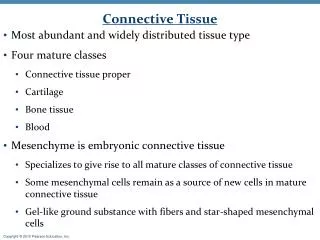
Morphologic forms • There are 4 morphologic forms seen in hemoflagellates: • Amastigote • Promastigote • Epimastigote • Trypomastigote -they can exist in two or more of the 4 morphologic forms depending on the species.
Kingdom: Protisata • Phylum: Sarcomastigophora • Class: Zoomastigophora • Order: Kimetoplastida • Family: Trypanosomatidae • Genus:Leishmania • Species:donovani , tropica, mexicana, braziliensis
Leismania sp. • Can cause: • Cutaneous leishmaniasis: a localized infection of the capillaries of the skin. • Mucocutaneous leishmaniasis: cause lesions of the skin and mucous membranes, specifically of the oral and nasal mucosa. • Visceral/sistemic leismaniasis: more generalized symptoms leading to enlargement of the internal organs, especially the liver, lymph nodes and spleen.
Leishmania sp. • Divided into 4 groups: 1) Leishmania tropica complex – Old World Cutaneous Leismaniasis. 2) Leishmania mexicana complex – New World Cutaneous Leishmaniasis. 3) Leishmania braziliensis complex – Mucocutaneous Laishmaniasis. 4) Leishmania donovani complex – Visceral leishmaniasis.
Stage of life • Only have 2 stages of life: • Amastigote • Promastigote
Amastigote • Size: 5 by 3µm • Shape: oval to round • Nucleus: One, eccentric. • Kinetoplast: Present, Consisting of dot-like blepharoplast, with small axoneme and prabasal body. • Flagellum: absent
Promastigote • Size: 9-15µm • Shape: long and slender. • Nucleus: one, central. • Kinetoplast: Anterior end of the organism, no undulating membrane. • Flagellum: Single, anterior free flagellum.
Leishmania tropica complex – Old World Cutaneous Leismaniasis. • L. tropica - mediterranean region, middle East, Armenia, Caspian region, Afghanistan, India and Kenya (particularly in urban areas)
Morphology • Cause a chronic disease: cutaneous leishmanisis. • Also known as Oriental sore, Delhi boil and dry or urban cutaneous leishmaniasis. • Characterized by: production of dry, raised, ulcerated lesions at bite sites. • Vectored by: tiny sandflies of the genera Phlebotomus.
Sandfly vs mosquito mosquito sandfly
Laboratory Diagnosis • Montenegro (leishmanin) skin test -delayed hypersensitivity reaction provoked by a suspension of killed leishmanial promastigotes administered intradermally. -local inflammatory reaction appears at the site of injection within 48-72 hours. • Microscopy examination • Isoenzyme studies • Molecular diagnostic technique- PCR • Serologic test – ex: indirect fluorescent antibody assay.
Leishmania braziliensis complex – Mucocutaneous Leishmaniasis • L. braziliensis – Mexico to Argentina
Morphology • Cause infections throughout the Americas from Mexico to Argentina. • The distinguishing feature of these infectious is the development of ulcers on or about the oral and nasal mucosa • L. braziliensis causes espundia.
Diagnosis • By demonstrating amastigotes of Leishmania in Giemsa stained smears or biopsy material from the edge of an active ulcer. • Cultivation. • Serologic test. • Montenegro skin test.
Leishmania donovani complex – Visceral leishmaniasis • L. donovani – India, Pakistan, Thailand, parts of Africa and the Peoples Republic of China. • L. infantum – Mediterranean area, Europe, Africa, the Near East, and parts of the former Soviet Union. • L. chagasi – Central and South America.
Morphology • Visceral leishmanisis also known as Kala Azar or dum-dum fever. • The most severe of the Leishmaniasis. • Generally a disease of juveniles and young adults. • Natural reservoir: rodents and dog. • In India, man appears to be the only mamalian reservoir. • L. donovani complex –parasitize the reticuloendothelial cells, viscerotropic, infected macrophages remaining fixed or disseminate throughout the body.
In the Mediterranean are, Europe, Africa, Soviet union – Phlebotomus sandfly remains the vector. Natural reservoir: domesticated dogs, canines and porcupines. • In the New World (Central and South America) – Lutzomiya sandfly remains the vector. Natural reservoir: Foxes, domestic dogs and cats.
Life cycle • Same with L. mexicana complex. • The infected mononuclear phagocytes do not remain confined to the skin or mucous membranes. • Parasitized macrophages are carried by the bloodstream to lymphoid tissue throughout the body especially to the spleen, liver and bone marrow. • Amastigotes multiply in great numbers in this tissues. • Vectors: Phlebotomus sandfly and Lutzomiya sandfly .
Diagnosis • Tissues Biopsy • Direct examination of stained smears. • Cultivation. • Serologic test. • Direct agglutination test (DAT). • Complement fixation test (CF). • Indirect fluorescence technique. • Molecular diagnostic technique. • Montenegro skin test (not reactive in people with active disease).
Promastigotes (left) and amastigotes (right) of Leishmania parasites. Parasites have been fixed in Giemsa stained.