Download
1 / 75
830 likes | 1.42k Views
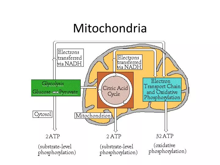
Mitochondria. Mitochondria. ER Targeting and Secretory Pathway. ER. Golgi. Vesicles. PM. Secretion. Lysosome. Endoplasmic Reticulum. Endoplasmic Reticulum. Two parts:. Endoplasmic Reticulum. Rough ER. Endoplasmic Reticulum. What happens in the ER ?. Targeting of Proteins to the ER.

E N D
ER Targeting and Secretory Pathway ER Golgi Vesicles PM Secretion Lysosome
Endoplasmic Reticulum • Two parts:
Endoplasmic Reticulum • Rough ER
Endoplasmic Reticulum • What happens in the ER?
Targeting of Proteins to the ER What proteins pass through ER? ER residents Other components of the secretory pathway Secreted proteins Proteins going to the plasma membrane
ER targeting is cotranslational Ribosomes free in cytosol Recruited to ER CotranslationalTranslocation
Targeting of Proteins to the ER PLAYERS In cytoplasm: Ribosome Peptide with signal sequence Signal Recognition Particle (SRP) In ER membrane: SRP Receptor Translocon Signal Peptidase BiP Oligosaccharide Transferase Cytoplasm OST BiP ER lumen
Targeting of Proteins to the ER • How are proteins targeted to the ER? • N-terminal signal sequence:
Signal sequence targets proteins to ER • In the cytosol: • Signal Recognition Particle (SRP): • Cytosolic ribonucleoprotein particle • Binds GTP • 6 polypeptides bound to a 300 nucleotide RNA molecule • RNA acts as a scaffold • SRP stops translation of mRNA upon binding • Binds to ER signal sequence and large ribosome subunit • Directs complex to the ER membrane so it can bind the receptor
Targeting of Proteins to the ER • On the ER membrane: • SRP receptor • Alpha and beta subunits; GTP bound in ER membrane • Binding of GTP strengthens the interaction between SRP and the SRP receptor • Translocon • Sec61α is a membrane protein with 10 membrane spanning α helicies • Gated and tightly regulated
ER protein targeting • Steps 1 and 2 • ER signal sequence emerges from the ribosome • SRP binds, stops translation
ER protein targeting • Step 3 • Complex is quickly targeted to the ER membrane and the SRP receptor
ER protein targeting ER protein targeting • Step 4 • Ribosome-cargo transferred to translocon • Channel opens • Signal sequence and polypeptide go into channel • Translation resumes
ER protein targeting ER protein targeting • Steps 5 and 6 • As the polypeptide elongates, it passes into lumen • Signal sequence is cleaved by signal peptidase and rapidly degrades
ER protein targeting ER protein targeting • Steps 7 and 8 • At the end of translation, ribosome is released • Protein is drawn into lumen • Transloconcloses • Protein folds 16
Translation drives translocation in ER lumen Translation drives translocation Signal sequence cleaved Ribosome released BiP (chaperone) - keeps protein unfolded Oligosaccharide transferase adds sugar
Microsomes • When cells are homogenized, ER forms microsomes • Contain ribosomes
Microsome allows in vitro study of membrane-bound processes Label secretory protein Homogenize ER to make microsomescontaining secretory protein OR make whole cell extracts without microsomes Treat with protease +/- detergent Run on gel to detect protein
- - - - Results + + + + Cell-free Microsomes protease detergent - - + + - - + + Secretory proteins translated in the presence of an ER are protected from protease. This means they are produced inside the ER
Protein Insertion into the ER • Membrane proteins for ER, Golgi and lysosomes are sythesizedin the ER • Topogenic sequences direct membrane insertion
Integral Membrane Classes • Differ by orientation and signal sequence
Orientation of protein in membrane important for its function What would happen if the Na/K ATPase pump were misoriented in the cell?
Two types of membrane signals Stop-Transfer Internal alpha-helical hydrophobic sequence of 22-25 amino acids Insufficient to target to translocon on its own; not a target of SRP Recognition of sequence causes translational stop and transfer of hydrophobic stretch to membrane Remaining translation continues in cytosol unless another signal sequence is encountered Signal Anchor Internal alpha-helical hydrophobic sequence of 22-25 amino acids with a stretch of positively charged amino acids to one side recognized by SRP and recruited to translocon Signal anchor amino acids are placed into membrane, but translation continues Location of translation is dictated by the orientation of the anchor (+++ amino acids)
Integral Membrane Classes Type I--Have a cleavable ER signal sequence
Type I • Type I proteins are targeted to the ER by SRP signal sequence mediated path • Signal sequence is cleaved in ER lumen • Stop Transfer Anchor Sequence • Sec61 • SRP and SRP receptor • N-term: • C-term:
Type I proteins contain N-term signal sequences and internal stop-transfer
Type II • Internal signal-anchor sequence (SA) • This stretch of amino acids tells the cell two things • SA sequence recruits SRP for targeting to the ER
Internal Signal Anchor Sequences are targeted to the ER by SRP Type II • 5’ end is translated in the cytosol • Translation of signal anchor sequence allows SRP to bind • SRP receptor • SA to translocon • Translation finishes
Type III • Two possible orientations for SA containing proteins: • The +++ charges will always be to the cytosolic side • In Type II, +++ on the N-terminal side of SA • In Type III, the +++ on the C-terminal side
Internal Signal Anchor Sequences are targeted to the ER by SRP Type III • Steps are similar to Type II • N-terminus is in the lumen • Due to the +++ charged amino acids on C-terminal side
Internal Signal Anchor Sequences are targeted to the ER by SRP Type II Type III • laskdfalksd
Transmembrane Sequences • Note the differences between the three types
Multipass Proteins use a combination Multipasstransmembrane proteins alternate between Stop-Transfer and Signal Anchor Sequences Odd: Even:
Multipassproteins- Type Iva and IVb • Type IVa
Multipass proteins • Type IVb
Secretory Pathway ER Golgi Secretory Vesicles Production, processing, and sorting of material bound for cell membrane components or secretion
Physical Apposition of Secretory Pathway Components Secretory vesicle Golgi ER-to-Golgi vesicles Rough ER Order of travel is critical for proper modifications
Golgi Complex • Most proteins leave ER within minutes • Where do they go? • How do they get there? • Why?
Vesicular Transport • Vesicles transport proteins to different target sites
Vesicular Transport from the ER • With proper folding correct modifications, proteins leave the ER • Transport vesicles
Target Vesicle Coat General Steps from ER to Golgi and back again cargo Donor Cargo accumulation G-protein binds to donor compartment Coat assembly and budding Coat release Target compartment fusion Vesicle components released
General Steps from ER to Golgi and back again Players G-protein that recruits Coat: Sar1 or ARF GEF in donor compartment Coat proteins Sorting signal sequence in cytosolic portion of transmembrane cargo G-protein that targets vesicle to correct destination: Rab Rab effector in target compartment Snares in vesicle and target compartment to aid in docking and fusion
General Steps from ER to Golgi and back again • Vesicle coat forms • Interacts with the cytosolic portion of membrane proteins • Coats provide curvature needed for budding • Small GTP-binding protein
COPII transport Overview • 5 steps
General Steps from ER to Golgi and back again • Step 1 • Sec12-GEF promotes GDP to GTP exchange in Sar1 • Causes conformation change • This drives polymerization of COPII
General Steps from ER to Golgi and back again • Step 2 • Sar1-GTP serves as a binding site • Coats provide the curvature • Membrane cargo is recruited by a binding site in their cytoplasmic portions