Download
1 / 40
440 likes | 469 Views
Learn about indirect immunological detection of Lyme disease through serological lab tests recommended by health organizations. Includes specifics of two-step diagnostics, ELISA, Western Blot methods, and key proteins targeted in testing.

E N D
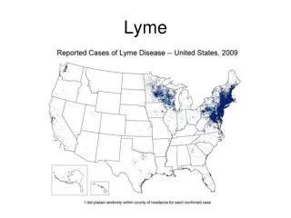
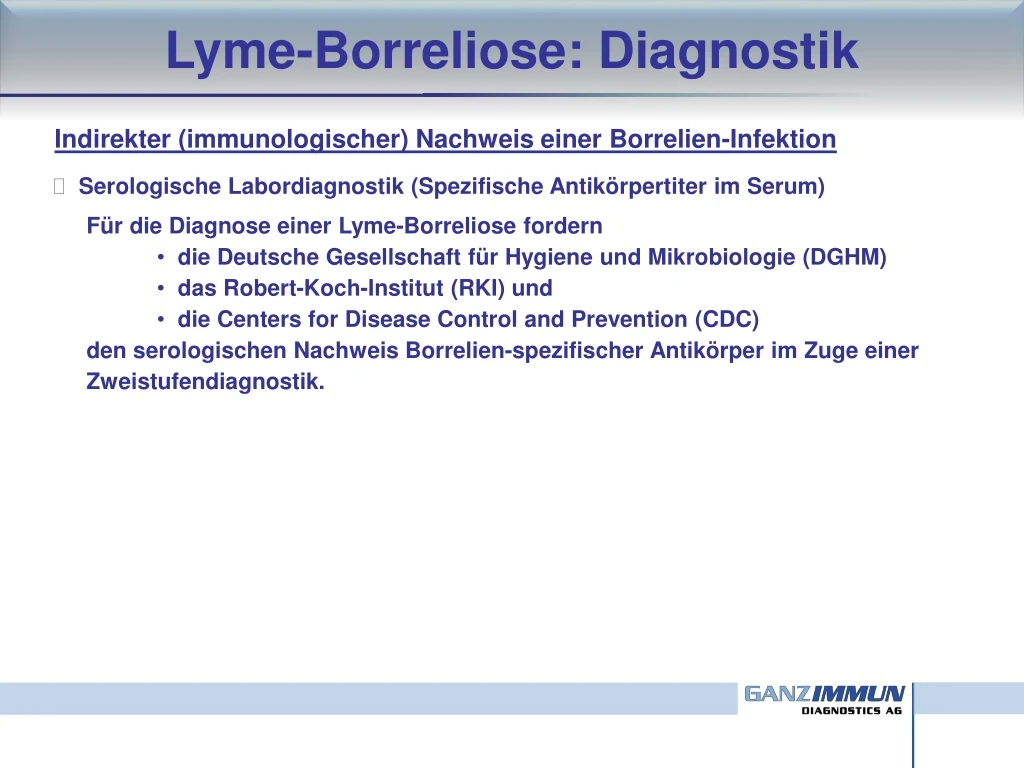
Lyme-Borreliose: Diagnostik Indirekter (immunologischer) Nachweis einer Borrelien-Infektion • Serologische Labordiagnostik (Spezifische Antikörpertiter im Serum) • Für die Diagnose einer Lyme-Borreliose fordern • die Deutsche Gesellschaft für Hygiene und Mikrobiologie (DGHM) • das Robert-Koch-Institut (RKI) und • die Centers forDiseaseControlandPrevention (CDC) • den serologischen Nachweis Borrelien-spezifischer Antikörper im Zuge einer • Zweistufendiagnostik.
Lyme-Borreliose: Diagnostik Indirekter (immunologischer) Nachweis einer Borrelien-Infektion • Serologische Labordiagnostik (Spezifische Antikörpertiter im Serum)
Lyme-Borreliose: Diagnostik Indirekter (immunologischer) Nachweis einer Borrelien-Infektion • Serologische Labordiagnostik (Spezifische Antikörpertiter im Serum) • Für die Diagnose einer Lyme-Borreliose fordern • die Deutsche Gesellschaft für Hygiene und Mikrobiologie (DGHM) • das Robert-Koch-Institut (RKI) und • die Centers forDiseaseControlandPrevention (CDC) • den serologischen Nachweis Borrelien-spezifischer Antikörper im Zuge einer • Zweistufendiagnostik. • 1. Stufe: Antikörper-Suchtest (IgM, IgG) positiv oder grenzwertig • 2. Stufe: Bestätigungstest (IgM, IgG) •
Lyme-Borreliose: Diagnostik Indirekter (immunologischer) Nachweis einer Borrelien-Infektion • Antikörper-Suchtest (IgM, IgG) • Testverfahren: ELISA (Enzym-gekoppelter Immun-Adsorptionstest) • Bestimmung von IgM und IgG gegen Proteine aus Extrakten von • B. burgdorferisensustrictu, • B. garinii und B. afzeliisowie • von IgG gegen rekombinantes • Oberflächenprotein VlsE • breites Antigenspektrum • hohe Sensitivität • unspezifische • Reaktionen unter • bestimmten • Bedingungen möglich
Lyme-Borreliose: Diagnostik Indirekter (immunologischer) Nachweis einer Borrelien-Infektion • Bestätigungstest (IgM, IgG) • Testverfahren: Western Blot • Farbbanden zeigen die Anwesenheit von • Antikörpern an • Bestimmung von IgM und IgG gegen definierte • Oberflächenproteine aus Borrelien (u.a. OspC und VlsE) Zeckendarm OspA+ OspC+ VlsE+ Speicheldrüsen der Zecke Hoviuset al. 2007. Tick–host–pathogen interactions in Lymeborreliosis. Trends Parasitol 23:434 Kenedyet al.2012. The role of Borreliaburgdorferi outer surface proteins. FEMS Immunol Med Microbiol 66:1
Lyme-Borreliose: Diagnostik Indirekter (immunologischer) Nachweis einer Borrelien-Infektion • Bestätigungstest (IgM, IgG) • Testverfahren: Western Blot • Farbbanden zeigen die Anwesenheit von • Antikörpern an • Bestimmung von IgM und IgG gegen definierte • Oberflächenproteine aus Borrelien (u.a. OspC und VlsE) VlsE = (variable major protein-like sequence, expressed) IgM IgG Schwan und Piesman. 2000. Temporal changes in outer surface proteins A and C of the Lyme disease-associated spirochete, Borreliaburgdorferi, during the chain of infection in ticks and mice. J ClinMicrobiol 38:382 Mülleret al. 2012. Evaluating frequency, diagnostic quality, and cost of Lyme borreliosis testing in Germany: a retrospective model analysis. Clin Dev Immunol ID595427.
Lyme-Borreliose: Diagnostik Indirekter (immunologischer) Nachweis einer Borrelien-Infektion • Bestätigungstest (IgM, IgG) • Testverfahren: Western Blot • Farbbanden zeigen die Anwesenheit von • Antikörpern an • Bestimmung von IgM und IgG gegen definierte • Oberflächenproteine aus Borrelien (u.a. OspC und VlsE) • hohe Spezifität und Sensitivität • Weiss et al. 1995. False positive seroreactivitytoBorreliaburgdorferi in • systemiclupuserythematosus: thevalueofimmunoblotanalysis. Lupus 4:131. In bis zu 40% der Patienten mit systemischem Lupus erythematosus (SLE) und anderen rheumatischen Erkrankungen wurde eine positive Seroreaktivität für Borrelien-spezifische Antikörper im ELISA nachge-wiesen. Die Spezifität der Serum-Antikörper konnte allerdings mittels Immunoblot-Technik nicht verifiziert werden.
Lyme-Borreliose: Diagnostik Indirekter (immunologischer) Nachweis einer Borrelien-Infektion • Bestätigungstest (IgM, IgG) • Testverfahren: Western Blot • Zuverlässige Differenzierung zwischen aktiver und abgelaufener Infektion oft schwierig oder gar nicht möglich
Lyme-Borreliose: Diagnostik Indirekter (immunologischer) Nachweis einer Borrelien-Infektion • Bestätigungstest (IgM, IgG) • Testverfahren: Western Blot • Farbbanden zeigen die Anwesenheit von • Antikörpern an • Bestimmung von IgM und IgG gegen definierte • Oberflächenproteine aus Borrelien (u.a. OspC und VlsE) • hohe Spezifität und Sensitivität • ca. 10% (bis 40%) seronegative Patienten mit Borreliose-Symptomatik
Lyme-Borreliose: Diagnostik Seronegative Patienten mit Borreliose-Symptomatik • Bestimmung der Borrelien-spezifischen Antikörper mittels ELISA in 23 Patienten mit diagnostiziertem EM und positivem Borrelien-Nachweis (in vitro-Kultur) in Hautbiopsien • 9 Patienten (41%) waren seronegativ • in 8 Patienten (35%) ließen sich nur IgM-Antikörper nachweisen • in 2 Patienten (8%) ließen sich nur IgG-Antikörper nachweisen • in 4 Patienten (16%) ließen sich sowohl IgM- als auch IgG-Antikörper • bestimmen
Lyme-Borreliose: Diagnostik Seronegative Patienten mit Borreliose-Symptomatik • Bestimmung der Borrelien-spezifischen Antikörper mittels ELISA in 22 Patienten mit diagnostizierter chronischer kutaner Borreliose • in allen 22 Patienten (100%) ließen sich IgG-Antikörper nachweisen • in nur 1 Patienten (5%) ließen sich zusätzlich auch IgM-Antikörper • bestimmen
Lyme-Borreliose: Diagnostik Mögliche Gründe für Seronegativität in Patienten mit Borreliose-Symptomatik • mangelnde Sensitivität des Detektionssystems • Serumnahme kurz nach der Infektion (< 4 Wochen nach Zeckenstich) • angeborene oder erworbene Immunsuppression beim Patienten • van Dop et al. 2013. Seronegative Lyme neuroborreliosis in a patient using rituximab. BMJ Case Rep. • 2012:007627 • van Dop et al. 2013. Seronegative Lyme neuroborreliosis in a patient using rituximab. BMJ Case Rep • 2012:007627 • Harrer et al. 2007. Seronegative Lyme neuroborreliosis in a patient on treatment for chronic lymphytic • leukemia. Infection 35:110. “Abstract We report on a patient who developed seronegative Lyme neuroborreliosis complicating chemotherapy for chronic lymphatic leukemia. After the fifth cycle of chemotherapy (FCR: fludarabine, cyclophosphamide, rituximab and prednisone) the 63-year-old patient developed night sweat, arthralgia in elbows, wrists, proximal interphalangeal joints (PIPs) and strong neuropathic pain in both legs, followed by paresthesia and hypesthesia in the feet, arms and face. ... Cerebrospinal fluid (CSF) analysis showed a lymphomononuclearpleocytosis and an elevation of protein. A broad diagnostic work-up was negative including a negative BorreliaIgG and IgM ELISA. The patient did not remember recent tick bites, but after specific questioning he recollected a transient erythema on his leg developing just before the start of the last cycle of chemotherapy. As the combination of neuropathic pain and arthralgia, the transient erythema and the lymphomononuclearpleocytosis raised the suspicion of Lyme neuroborreliosis, the patient was treated for 3 weeks with ceftriaxone. On therapy all symptoms resolved and CRP normalized. Retrospective PCR analysis of a CSF sample confirmed the clinical diagnosis by detecting Borreliagarinii DNA. This case demonstrates that in immunosuppressed patients borrelial serology may be negative and that additional diagnostic approaches (…) may be needed to demonstrate borrelial infection.” “Abstract We report on a patient who developed seronegative Lyme neuroborreliosis complicating chemotherapy for chronic lymphatic leukemia. After the fifth cycle of chemotherapy (FCR: fludarabine, cyclophosphamide, rituximab and prednisone) the 63-year-old patient developed night sweat, arthralgia in elbows, wrists, proximal interphalangeal joints (PIPs) and strong neuropathic pain in both legs, followed by paresthesia and hypesthesia in the feet, arms and face. ... Cerebrospinal fluid (CSF) analysis showed a lymphomononuclearpleocytosis and an elevation of protein. A broad diagnostic work-up was negative including a negative BorreliaIgG and IgM ELISA. The patient did not remember recent tick bites, but after specific questioning he recollected a transient erythema on his leg developing just before the start of the last cycle of chemotherapy. As the combination of neuropathic pain and arthralgia, the transient erythema and the lymphomononuclearpleocytosis raised the suspicion of Lyme neuroborreliosis, the patient was treated for 3 weeks with ceftriaxone. On therapy all symptoms resolved and CRP normalized. Retrospective PCR analysis of a CSF sample confirmed the clinical diagnosis by detecting Borreliagarinii DNA. This case demonstrates that in immunosuppressed patients borrelial serology may be negative and that additional diagnostic approaches (…) may be needed to demonstrate borrelial infection.”
Lyme-Borreliose: Diagnostik Mögliche Gründe für Seronegativität in Patienten mit Borreliose-Symptomatik • mangelnde Sensitivität des Detektionssystems • Serumnahme kurz nach der Infektion (< 4 Wochen nach Zeckenstich) • angeborene oder erworbene Immunsuppression beim Patienten • Immunkomplexbildung Verhinderung der Detektion von freien Antikörpern • Schutzer et al. 1990. Sequestration of antibody to Borreliaburgdorferi in immune complexes in • seronegative Lyme disease. Lancet 335:312. • Schutzer et al. 1999. Borreliaburgdorferi-specific immune complexes in acute Lyme disease. • JAMA 282:1942. • Brunner. 2001. New method for detection of Borreliaburgdorferi antigen complexed to antibody in • seronegative Lyme disease. J Immunol Meth 249:185. • Marques et al. 2005. Detection of immune complexes is not independent of detection of antibodies • in Lyme disease patients and does not confirm active infection with Borreliaburgdorferi. • ClinDiagn Lab Immunol 12:1036.
Lyme-Borreliose: Diagnostik Mögliche Gründe für Seronegativität in Patienten mit Borreliose-Symptomatik • mangelnde Sensitivität des Detektionssystems • Serumnahme kurz nach der Infektion (< 4 Wochen nach Zeckenstich) • angeborene oder erworbene Immunsuppression beim Patienten • Immunkomplexbildung Verhinderung der Detektion von freien Antikörpern • Assoziation der Produktion Borrelien-spezifischer Antikörper mit bestimmten • HLA-Allelen • HLA-DR6/-DR7 ist assoziiert mit Antikörperproduktion • Seropositive Patienten: 16/22 (72,7%) • Seronegative Patienten: 2/18 (11,1%) • Kontrolle (gesunde Probanden): 13/26 (50,0%) • HLA-DR1 ist assoziiert mit dem Fehlen einer Antikörperproduktion • Seropositive Patienten: 1/22 (4,5%) • Seronegative Patienten: 7/18 (38,9%) • Kontrolle (gesunde Probanden): 3/26 (11,5%) • Wang und Hilton. 2001. Contribution of HLA alleles in the regulation of antibody production in Lyme • disease. Front Biosci 6:B10
Lyme-Borreliose: Diagnostik Mögliche Gründe für Seronegativität in Patienten mit Borreliose-Symptomatik • mangelnde Sensitivität des Detektionssystems • Serumnahme kurz nach der Infektion (< 4 Wochen nach Zeckenstich) • angeborene oder erworbene Immunsuppression beim Patienten • Immunkomplexbildung Verhinderung der Detektion von freien Antikörpern • Assoziation der Produktion Borrelien-spezifischer Antikörper mit bestimmten • HLA-Allelen • Frühe antibiotische Behandlung vermindert oder verhindert Antikörperbildung • Dattwyler et al. 1988. Seronegative Lyme disease. Dissociation of specific T- and B-lymphocyte • responses to Borreliaburgdorferi. N Engl J Med 319:1441 Abstract We studied 17 patients who had presented with acute Lyme disease and received prompt treatment with oral antibiotics, but in whom chronic Lyme disease subsequently developed. Although these patients had clinically active disease, none had diagnostic levels of antibodies to B. burgdorferi on either a standard enzyme-linked immunosorbent assay or immunofluorescence assay. On Western blot analysis, the level of immunoglobulin reactivity against B. burgdorferi in serum from these patients was no greater than that in serum from normal controls.
Lyme-Borreliose: Diagnostik Mögliche Gründe für Seronegativität in Patienten mit Borreliose-Symptomatik • mangelnde Sensitivität des Detektionssystems • Serumnahme kurz nach der Infektion (< 4 Wochen nach Zeckenstich) • angeborene oder erworbene Immunsuppression beim Patienten • Immunkomplexbildung Verhinderung der Detektion von freien Antikörpern • Assoziation der Produktion Borrelien-spezifischer Antikörper mit bestimmten • HLA-Allelen • Frühe antibiotische Behandlung vermindert oder verhindert Antikörperbildung Zelluläre Labordiagnostik (Bestimmung der spezifischen T-Zell-Reaktivität durch Funktionstestungen)
Lyme-Borreliose: Diagnostik Indirekter (immunologischer) Nachweis einer Borrelien-Infektion Abstract We studied 17 patients who had presented with acute Lyme disease and received prompt treatment with oral antibiotics, but in whom chronic Lyme disease subsequently developed. Although these patients had clinically active disease, none had diagnostic levels of antibodies to B. burgdorferi on either a standard enzyme-linked immunosorbent assay or immunofluorescence assay. On Western blot analysis, the level of immunoglobulin reactivity against B. burgdorferi in serum from these patients was no greater than that in serum from normal controls.The patients had a vigorous T-cell proliferative response to whole B. burgdorferi, with a mean ( +/- SEM) stimulation index of 17.8 +/- 3.3, similar to that (15.8 +/- 3.2) in 18 patients with chronic Lyme disease who had detectable antibodies. The T-cell response of both groups was greater than that of a control • Dattwyler et al. 1988. Seronegative Lyme disease. Dissociation of specific T- and B-lymphocyte • responses to Borreliaburgdorferi. N Engl J Med 319:1441 Abstract We studied 17 patients who had presented with acute Lyme disease and received prompt treatment with oral antibiotics, but in whom chronic Lyme disease subsequently developed. Although these patients had clinically active disease, none had diagnostic levels of antibodies to B. burgdorferi on either a standard enzyme-linked immunosorbent assay or immunofluorescence assay. On Western blot analysis, the level of immunoglobulin reactivity against B. burgdorferi in serum from these patients was no greater than that in serum from normal controls.The patients had a vigorous T-cell proliferative response to whole B. burgdorferi, with a mean ( +/- SEM) stimulation index of 17.8 +/- 3.3, similar to that (15.8 +/- 3.2) in 18 patients with chronic Lyme disease who had detectable antibodies. The T-cell response of both groups was greater than that of a control group of healthy subjects (3.1 +/- 0.5; P less than 0.001). We conclude that the presence of chronic Lyme disease cannot be excluded by the absence of antibodies against B. burgdorferi and that a specific T-cell blastogenic response to B. burgdorferi is evidence of infection in seronegative patients with clinical indications of chronic Lyme disease.” group of healthy subjects (3.1 +/- 0.5; P less than 0.001). We conclude that the presence of chronic Lyme disease cannot be excluded by the absence of antibodies against B. burgdorferi and that a specific T-cell blastogenic response to B. burgdorferi is evidence of infection in seronegative patients with clinical indications of chronic Lyme disease.”
Lyme-Borreliose: Diagnostik Indirekter (immunologischer) Nachweis einer Borrelien-Infektion • Lymphozytentransformationstest (LTT) • Prinzip: Bestimmung der Anwesenheit/Frequenz Borrelien-spezifischer • T-Lymphozyten im Blut als Indikator für eine vorausgegangene Infektion • Methode: Messung der Proliferation (Zellteilung) von Gedächtnis-T-Zellen • durch Quantifizierung des Einbaus eines radioaktiv markierten Nukleotids • (3H-Thymidin) in die DNA von Lymphozyten, die mit Borrelien-Antigenen • langzeitkultiviert (5 bis 6 Tage) werden. Lymphozyten-Aktivierung Transformation (Blastenbildung) Proliferation (Zellvermehrung) Han et al.2007. Nature Reviews Microbiology 4:95
Lyme-Borreliose: Diagnostik Indirekter (immunologischer) Nachweis einer Borrelien-Infektion • Lymphozytentransformationstest (LTT) • Prinzip: Bestimmung der Anwesenheit/Frequenz Borrelien-spezifischer • T-Lymphozyten im Blut als Indikator für eine vorausgegangene Infektion • Methode: Messung der Proliferation (Zellteilung) von Gedächtnis-T-Zellen • durch Quantifizierung des Einbaus eines radioaktiv markierten Nukleotids • (3H-Thymidin) in die DNA von Lymphozyten, die mit Borrelien-Antigenen • langzeitkultiviert (5 bis 6 Tage) werden. • Synonyme Bezeichnungen: • •Lymphozyten-Aktivierungstest (LAT) • T-Zell-Proliferationstest • LTT-MELISA (MEmoryLymphocyteImmunoStimulationAssay) • 3HT-Memory-Test
Lyme-Borreliose: Diagnostik Indirekter (immunologischer) Nachweis einer Borrelien-Infektion Zellkultur: Stimulation der mononukleären Zellen mit Borrelien-Antigenen für 5 bis 6 Tage • Lymphozytentransformationstest (LTT) unstimuliert stimuliert Mit freundlicher Genehmigung von Dr. Verena Raker (Universitätsmedizin Mainz) Proliferation in stimulierter Kultur [cpm] Proliferation in unstimulierter Kultur [cpm] SI > Schwellenwert Reaktion positiv Stimulationsindex SI = -------------------------------------------------------
Lyme-Borreliose: Diagnostik Indirekter (immunologischer) Nachweis einer Borrelien-Infektion • Prinzip: Bestimmung der Anwesenheit/Frequenz Borrelien-spezifischer • T-Lymphozyten im Blut als Indikator für eine vorausgegangene Infektion • Lymphozytentransformationstest (LTT) • Methode: Messung der Proliferation (Zellteilung) von Gedächtnis-T-Zellen • durch Quantifizierung des Einbaus eines radioaktiv markierten Nukleotids • (3H-Thymidin) in die DNA von Lymphozyten, die mit Borrelien-Antigenen • langzeitkultiviert (5 bis 6 Tage) werden. • Synonyme Bezeichnungen: • •Lymphozyten-Aktivierungstest (LAT) • T-Zell-Proliferationstest • LTT-MELISA (MEmoryLymphocyteImmunoStimulationAssay) • 3HT-Memory-Test • Die Eignung des LTT als diagnostisches Verfahren zur Bestätigung einer • Borreliose ist labormedizinisch umstritten. Vor allen Dingen in älteren • Studien (vor 2000) werden Sensitivität und/oder Spezifität des LTT als • nicht ausreichend für eine valide Bewertung der Ergebnisse beurteilt.
Lyme-Borreliose: Diagnostik Indirekter (immunologischer) Nachweis einer Borrelien-Infektion • Lymphozytentransformationstest (LTT) OBJECTIVE: To determine the sensitivity and specificity of the T-cell proliferative assay as a diagnostic test in Lyme disease. DESIGN: Cross-sectional study of patients with Lyme arthritis or chronic neuroborreliosis who had a history of erythemamigrans, positive antibody responses to Borreliaburgdorferi by enzyme-linked immunosorbent assay (ELISA), or both; patients with other diseases; and healthy subjects. PATIENTS: Forty-two of the 67 patients with active Lyme arthritis or chronic neuroborreliosis who were seen during the study period; 16 patients with inactive late Lyme disease; 77 patients with other rheumatologic or neurologic diseases; 9 workers from the Borrelia laboratory; and 9 healthy subjects. MEASUREMENTS AND MAIN RESULTS: Nineteen of 42 patients with Lyme arthritis or chronic neuroborreliosis and 4 of 77 patients with other diseases had positive T-cell proliferative responses to B. burgdorferi antigens. The sensitivity of the proliferative assay was 45% (95% Cl, 30% to 60%) and the specificity was 95% (95% Cl, 87% to 99%). Twelve of 27 patients with active Lyme arthritis, 7 of 15 patients with chronic neuroborreliosis, 4 of 16 patients with inactive Lyme disease, 4 of 9 healthy Borrelia laboratory workers, and 0 of 9 healthy subjects had positive responses. Three of five patients with Lyme disease who had negative or indeterminant antibody responses by ELISA had positive T-cell proliferative responses. CONCLUSION: The T-cell proliferative assay may be a helpful diagnostic test in the small subset of patients with late Lyme disease who have negative or indeterminant antibody responses by ELISA. OBJECTIVE: To determine the sensitivity and specificity of the T-cell proliferative assay as a diagnostic test in Lyme disease. DESIGN: Cross-sectional study of patients with Lyme arthritis or chronic neuroborreliosis who had a history of erythemamigrans, positive antibody responses to Borreliaburgdorferi by enzyme-linked immunosorbent assay (ELISA), or both; patients with other diseases; and healthy subjects. PATIENTS: Forty-two of the 67 patients with active Lyme arthritis or chronic neuroborreliosis who were seen during the study period; 16 patients with inactive late Lyme disease; 77 patients with other rheumatologic or neurologic diseases; 9 workers from the Borrelia laboratory; and 9 healthy subjects. MEASUREMENTS AND MAIN RESULTS: Nineteen of 42 patients with Lyme arthritis or chronic neuroborreliosis and 4 of 77 patients with other diseases had positive T-cell proliferative responses to B. burgdorferi antigens. The sensitivity of the proliferative assay was 45% (95% Cl, 30% to 60%) and the specificity was 95% (95% Cl, 87% to 99%). Twelve of 27 patients with active Lyme arthritis, 7 of 15 patients with chronic neuroborreliosis, 4 of 16 patients with inactive Lyme disease, 4 of 9 healthy Borrelia laboratory workers, and 0 of 9 healthy subjects had positive responses. Three of five patients with Lyme disease who had negative or indeterminant antibody responses by ELISA had positive T-cell proliferative responses. CONCLUSION: The T-cell proliferative assay may be a helpful diagnostic test in the small subset of patients with late Lyme disease who have negative or indeterminant antibody responses by ELISA. • Dressler et al. 1991. The T-cell proliferative assay in the diagnosis of Lyme disease. • Ann Intern Med 115:533. OBJECTIVE: To determine the sensitivity and specificity of the T-cell proliferative assay as a diagnostic test in Lyme disease. DESIGN: Cross-sectional study of patients with Lyme arthritis or chronic neuroborreliosis who had a history of erythemamigrans, positive antibody responses to Borreliaburgdorferi by enzyme-linked immunosorbent assay (ELISA), or both; patients with other diseases; and healthy subjects. PATIENTS: Forty-two of the 67 patients with active Lyme arthritis or chronic neuroborreliosis who were seen during the study period; 16 patients with inactive late Lyme disease; 77 patients with other rheumatologic or neurologic diseases; 9 workers from the Borrelia laboratory; and 9 healthy subjects. MEASUREMENTS AND MAIN RESULTS: Nineteen of 42 patients with Lyme arthritis or chronic neuroborreliosis and 4 of 77 patients with other diseases had positive T-cell proliferative responses to B. burgdorferi antigens. The sensitivity of the proliferative assay was 45% (95% Cl, 30% to 60%) and the specificity was 95% (95% Cl, 87% to 99%). Twelve of 27 patients with active Lyme arthritis, 7 of 15 patients with chronic neuroborreliosis, 4 of 16 patients with inactive Lyme disease, 4 of 9 healthy Borrelia laboratory workers, and 0 of 9 healthy subjects had positive responses. Three of five patients with Lyme disease who had negative or indeterminant antibody responses by ELISA had positive T-cell proliferative responses. CONCLUSION: The T-cell proliferative assay may be a helpful diagnostic test in the small subset of patients with late Lyme disease who have negative or indeterminant antibody responses by ELISA.
Lyme-Borreliose: Diagnostik Indirekter (immunologischer) Nachweis einer Borrelien-Infektion • Lymphozytentransformationstest (LTT) BACKGROUND AND DESIGN: ... We studied the lymphoproliferative response of peripheral blood mononuclear cells to B burgdorferi antigen from 99 patients (25 with erythemamigrans, 16 with acrodermatitischronicaatrophicans, 13 with lymphadenosisbenigna cutis, and 45 with localized scleroderma) and 21 control subjects. ... RESULTS: The 21 healthy seronegative controls had an SI of 3.3 +/- 2.0 (mean +/- SD). Compared with that of control subjects, the SIs were significantly elevated in patients with erythemamigrans (9.8 +/- 9.1), acrodermatitischronicaatrophicans (11.8 +/- 8.2), and lymphadenosisbenigna cutis (7.2 +/- 6.2). ... Elevated titers of antibodies to B burgdorferi were present in six (24%) of 25 patients with erythemamigrans, five (38%) of 13 patients with lymphadenosisbenigna cutis, and 13 (29%) of 45 patients with localized scleroderma. All 16 patients with acrodermatitischronicaatrophicans had markedly elevated antibody titers. CONCLUSIONS: Our findings show that a significant lymphoproliferative response to B burgdorferi occurs in the majority of patients with cutaneous manifestations of Lyme borreliosis. The lymphocyte proliferation assay may be of diagnostic value in patients in whom Lyme borreliosis is strongly clinically suspected and who have nondiagnostic levels of antibodies against B burgdorferi. • Buechner et al. 1995. Lymphoproliferative responses to Borreliaburgdorferi in patients with erythema • migrans, acrodermatitischronicaatrophicans, lymphadenosisbenigna cutis, and morphea. • Arch Dermatol 131:673. BACKGROUND AND DESIGN: ... We studied the lymphoproliferative response of peripheral blood mononuclear cells to B burgdorferi antigen from 99 patients (25 with erythemamigrans, 16 with acrodermatitischronicaatrophicans, 13 with lymphadenosisbenigna cutis, and 45 with localized scleroderma) and 21 control subjects. ... RESULTS: The 21 healthy seronegative controls had an SI of 3.3 +/- 2.0 (mean +/- SD). Compared with that of control subjects, the SIs were significantly elevated in patients with erythemamigrans (9.8 +/- 9.1), acrodermatitischronicaatrophicans (11.8 +/- 8.2), and lymphadenosisbenigna cutis (7.2 +/- 6.2). ... Elevated titers of antibodies to B burgdorferi were present in six (24%) of 25 patients with erythemamigrans, five (38%) of 13 patients with lymphadenosisbenigna cutis, and 13 (29%) of 45 patients with localized scleroderma. All 16 patients with acrodermatitischronicaatrophicans had markedly elevated antibody titers. CONCLUSIONS: Our findings show that a significant lymphoproliferative response to B burgdorferi occurs in the majority of patients with cutaneous manifestations of Lyme borreliosis. The lymphocyte proliferation assay may be of diagnostic value in patients in whom Lyme borreliosis is strongly clinically suspected and who have nondiagnostic levels of antibodies against B burgdorferi.
Lyme-Borreliose: Diagnostik Indirekter (immunologischer) Nachweis einer Borrelien-Infektion • Lymphozytentransformationstest (LTT) • Breier et al. 1995. Lymphocyteproliferationtest in cutaneousmanifestationsofLymeborreliosis. • Wien Med Wochenschr 145:170 • Breier et al. 1995. Lymphocyteproliferationtest in cutaneousmanifestationsofLymeborreliosis. • Wien Med Wochenschr 145:170. ABSTRACT The humoral immunoreactivity in Lymeborreliosisis a well characterizedparameterforestablishingthediagnosisof a Borreliaburgdorferi (Bb) infection. SincepatientswithseronegativeLymeborreliosishavebeendescribed, lymphocyteproliferationtestsmaybeusedfordetectingpatientswhoonlydevelop a cellularimmunoreactivityagainstBborganisms. Weperformed a thelymphocyteproliferationassays in order todeterminethecellularimmunoreactivitytothesespirochetes in givenpatientswithhistologicallyconfirmeddiagnosisoferythemamigrans (EM), acrodermatitischronicaatrophicans (ACA), andforcontrolpurposespatientswithLymedisease non associateddermatoses (NLDH) andhealthyvolunteers (G). … . Wedetectedelevatedcellularimmunoreactivityofperipheralmononuclearcells in 33% EM-patients (3/9) and in 23% (3/13) ACA patientstested. Patientstestedbeforeand after antibiotictherapyshowed a significantdecreaseofstimulationindex after antimicrobialtreatment (p < 0.05). Patients, whoaremostlyseronegativeattheonsetofcutaneouseruptionexhibit in a thirdofpatients an elevatedcellular immune responsetoBb. Thereforelymphocyteproliferationassayscanberecommendedas an additional testsystem in caseof lack ofserologicalresponse. The significantdecreaseofstimulationindex after antimicrobialtherapyindicatesfordownregulationofthecellular immune responsetothesespirochetesinvestigated, andcountsforthespecificityofthistestsystem. ABSTRACT The humoral immunoreactivity in Lymeborreliosisis a well characterizedparameterforestablishingthediagnosisof a Borreliaburgdorferi (Bb) infection. SincepatientswithseronegativeLymeborreliosishavebeendescribed, lymphocyteproliferationtestsmaybeusedfordetectingpatientswhoonlydevelop a cellularimmunoreactivityagainstBborganisms. Weperformed a thelymphocyteproliferationassays in order todeterminethecellularimmunoreactivitytothesespirochetes in givenpatientswithhistologicallyconfirmeddiagnosisoferythemamigrans (EM), acrodermatitischronicaatrophicans (ACA), andforcontrolpurposespatientswithLymedisease non associateddermatoses (NLDH) andhealthyvolunteers (G). … . Wedetectedelevatedcellularimmunoreactivityofperipheralmononuclearcells in 33% EM-patients (3/9) and in 23% (3/13) ACA patientstested. Patientstestedbeforeand after antibiotictherapyshowed a significantdecreaseofstimulationindex after antimicrobialtreatment (p < 0.05). Patients, whoaremostlyseronegativeattheonsetofcutaneouseruptionexhibit in a thirdofpatients an elevatedcellular immune responsetoBb. Thereforelymphocyteproliferationassayscanberecommendedas an additional testsystem in caseof lack ofserologicalresponse. The significantdecreaseofstimulationindex after antimicrobialtherapyindicatesfordownregulationofthecellular immune responsetothesespirochetesinvestigated, andcountsforthespecificityofthistestsystem.
Lyme-Borreliose: Diagnostik Indirekter (immunologischer) Nachweis einer Borrelien-Infektion • Lymphozytentransformationstest (LTT) ABSTRACT To assess the contribution of the lymphocyte proliferation assay in response to borrelial antigens to establishing a diagnosis of Lyme arthritis (LA) the response to two strains of Borreliaburgdorferi was tested in peripheral blood lymphocytes of 103 children and adolescents with arthritis, among them 55 with LA and 48 control patients. Patients with LA had a significantly higher response to borrelial antigens than control patients. However, there were several patients with false positive and false negative test results. Specificity and sensitivity of the test were 78% and 77%. In patients with LA the test may turn positive after antibiotic therapy and remain positive for up to 19 months after the disappearance of arthritis. The test does not aid in prognosis or follow up. In one patient with seronegative LA specific lymphocyte proliferation and polymerase chain reaction for borrelialfla sequences in urine were positive. CONCLUSION Rarely the lymphocyte proliferation assay may aid in finding the correct diagnosis when clinical presentation and anti-borrelial serology do not match. ABSTRACT To assess the contribution of the lymphocyte proliferation assay in response to borrelial antigens to establishing a diagnosis of Lyme arthritis (LA) the response to two strains of Borreliaburgdorferi was tested in peripheral blood lymphocytes of 103 children and adolescents with arthritis, among them 55 with LA and 48 control patients. Patients with LA had a significantly higher response to borrelial antigens than control patients. However, there were several patients with false positive and false negative test results. Specificity and sensitivity of the test were 78% and 77%. In patients with LA the test may turn positive after antibiotic therapy and remain positive for up to 19 months after the disappearance of arthritis. The test does not aid in prognosis or follow up. In one patient with seronegative LA specific lymphocyte proliferation and polymerase chain reaction for borrelialfla sequences in urine were positive. CONCLUSION Rarely the lymphocyte proliferation assay may aid in finding the correct diagnosis when clinical presentation and anti-borrelial serology do not match. • Huppertz et al. 1996. Lymphoproliferative responses to Borreliaburgdorferi in the diagnosis of Lyme • arthritis in children and adolescents. Eur J Pediatr 155:297. ABSTRACT To assess the contribution of the lymphocyte proliferation assay in response to borrelial antigens to establishing a diagnosis of Lyme arthritis (LA) the response to two strains of Borreliaburgdorferi was tested in peripheral blood lymphocytes of 103 children and adolescents with arthritis, among them 55 with LA and 48 control patients. Patients with LA had a significantly higher response to borrelial antigens than control patients. However, there were several patients with false positive and false negative test results. Specificity and sensitivity of the test were 78% and 77%. In patients with LA the test may turn positive after antibiotic therapy and remain positive for up to 19 months after the disappearance of arthritis. The test does not aid in prognosis or follow up. In one patient with seronegative LA specific lymphocyte proliferation and polymerase chain reaction for borrelialfla sequences in urine were positive. CONCLUSION Rarely the lymphocyte proliferation assay may aid in finding the correct diagnosis when clinical presentation and anti-borrelial serology do not match.
Lyme-Borreliose: Diagnostik Indirekter (immunologischer) Nachweis einer Borrelien-Infektion • Lymphozytentransformationstest (LTT) ABSTRACT Humoral immune responses to Borreliaburgdorferi (Bb) have been reported to occur in certain patients with circumscribed scleroderma (CS) (morphoea). Together with the isolation of spirochaetes from CS skin biopsies, this finding was taken to suggest Bb as the aetiological agent of CS. ... For this purpose, peripheral blood mononuclear cells from CS patients and, as controls, from patients with various manifestations of LB, and from healthy volunteers without any evidence of Bb infection, were exposed to Bb organisms for 5 days and then assayed for DNA synthesis. Stimulation indices (SI) > 10 were scored positive. By performing lymphocyte proliferation tests we found: (i) that not only patients with various manifestations of LB but also a considerable percentage of seropositive (five of 13 = 38%) and seronegative (six of 26 = 23%) CS patients exhibit an elevated Bb-induced lymphocyte proliferation; (ii) that the magnitude of the cellular response seen in CS patients is comparable to that encountered in patients with established Bb manifestations; and (iii) that, within a given patient, antibiotic therapy can result in a significant reduction of this response. These results support a causative role of Bb in at least some CS patients. Bb-induced lymphocyte responses were also seen in both seropositive and seronegativeerythemachronicummigrans patients. These findings show that the pattern of Bb-specific immune responses is more complex than previously thought, and underscore the importance of lymphocyte function assays in evaluating the diagnosis of potential Bb infection in seronegative patients. • Breier et al. 1996. Lymphoproliferative responses to Borreliaburgdorferi in circumscribed • scleroderma. Br J Dermatol 134:285. ABSTRACT Humoral immune responses to Borreliaburgdorferi (Bb) have been reported to occur in certain patients with circumscribed scleroderma (CS) (morphoea). Together with the isolation of spirochaetes from CS skin biopsies, this finding was taken to suggest Bb as the aetiological agent of CS. ... For this purpose, peripheral blood mononuclear cells from CS patients and, as controls, from patients with various manifestations of LB, and from healthy volunteers without any evidence of Bb infection, were exposed to Bb organisms for 5 days and then assayed for DNA synthesis. Stimulation indices (SI) > 10 were scored positive. By performing lymphocyte proliferation tests we found: (i) that not only patients with various manifestations of LB but also a considerable percentage of seropositive (five of 13 = 38%) and seronegative (six of 26 = 23%) CS patients exhibit an elevated Bb-induced lymphocyte proliferation; (ii) that the magnitude of the cellular response seen in CS patients is comparable to that encountered in patients with established Bb manifestations; and (iii) that, within a given patient, antibiotic therapy can result in a significant reduction of this response. These results support a causative role of Bb in at least some CS patients. Bb-induced lymphocyte responses were also seen in both seropositive and seronegativeerythemachronicummigrans patients. These findings show that the pattern of Bb-specific immune responses is more complex than previously thought, and underscore the importance of lymphocyte function assays in evaluating the diagnosis of potential Bb infection in seronegative patients.
Lyme-Borreliose: Diagnostik Indirekter (immunologischer) Nachweis einer Borrelien-Infektion • Lymphozytentransformationstest (LTT) ABSTRACT We determined cellular and humoral immune responses to Borreliaburgdorferilysate and to recombinant flagellin (FlaB), OspC, and OspAin acute- and convalescent-phase samples from 39 culture-positive patients with erythemamigrans and in 20 healthy control subjects. During the acute illness, a median of 4 days after the onset of erythemamigrans, 51% of the patients had proliferative cellular responses and 72% had antibody responses to at least one of the borrelial antigens tested. During convalescence, at the conclusion of antibiotic therapy, 64% of the patients had proliferative cellular reactivity and 95% had antibody reactivity with at least one of the spirochetal antigens tested. In both acute- and convalescent-phase samples, cellular immune responses were found as frequently to OspA as to OspC and FlaB. Although antibody responses were also frequently seen to OspC and FlaB, only a few patients had marginal antibody reactivity with OspA. The percentage of patients with proliferative responses was similar in those with clinical evidence of localized or disseminated infection, whereas humoral reactivity was found more often in those with disseminated disease. We conclude that cellular and humoral responses to B. burgdorferi antigens are often found among patients with early Lyme disease. In contrast with the other antigens tested, cellular but not humoral reactivity was often found with OspA. • Vaz et al. 2001. Cellular and humoral immune responses to Borreliaburgdorferi antigens in patients • with culture-positive early Lyme disease. Infect Immun 69:7437. ABSTRACT We determined cellular and humoral immune responses to Borreliaburgdorferilysate and to recombinant flagellin (FlaB), OspC, and OspA in acute- and convalescent-phase samples from 39 culture-positive patients with erythemamigrans and in 20 healthy control subjects. During the acute illness, a median of 4 days after the onset of erythemamigrans, 51% of the patients had proliferative cellular responses and 72% had antibody responses to at least one of the borrelial antigens tested. During convalescence, at the conclusion of antibiotic therapy, 64% of the patients had proliferative cellular reactivity and 95% had antibody reactivity with at least one of the spirochetal antigens tested. In both acute- and convalescent-phase samples, cellular immune responses were found as frequently to OspA as to OspC and FlaB. Although antibody responses were also frequently seen to OspC and FlaB, only a few patients had marginal antibody reactivity with OspA. The percentage of patients with proliferative responses was similar in those with clinical evidence of localized or disseminated infection, whereas humoral reactivity was found more often in those with disseminated disease. We conclude that cellular and humoral responses to B. burgdorferi antigens are often found among patients with early Lyme disease. In contrast with the other antigens tested, cellular but not humoral reactivity was often found with OspA.
Lyme-Borreliose: Diagnostik Indirekter (immunologischer) Nachweis einer Borrelien-Infektion • Lymphozytentransformationstest (LTT) • Vaz et al. 2001. Cellular and humoral immune responses to Borreliaburgdorferi antigens in patients • with culture-positive early Lyme disease. Infect Immun 69:7437. Results Cellular immune responses to B. burgdorferi did not correlate with humoral immune reactivity. Moreover, there were patients with weak cellular reactivity and strong humoral responses or vice versa (data not shown). Overall, cellular reactivity with OspA was as frequent and as strong as the cellular responses to FlaB and OspC.
Lyme-Borreliose: Diagnostik Indirekter (immunologischer) Nachweis einer Borrelien-Infektion • Lymphozytentransformationstest (LTT) ABSTRACT The measurement of the proliferative response of primed T cells to an antigenic stimulus (lymphocyte transformation assay: LTT) is commonly used for determining T cell immune responsiveness. However, the ratio between the spontaneous and the antigen-triggered response (stimulation index) is frequently quite low (<3-5) making the interpretation difficult. We modified the assay by the addition of interferon-alpha and the use of fresh autologous serum instead of human AB pool serum. These measures significantly enhanced the stimulation index following stimulation with tetanus toxoid, Candida albicans and tick-borne encephalitis (TBE) viral antigen in studies of sensitized patients. There was no concomitant increase in false positive results. Kinetic studies showed a reduced nonspecific background proliferation of non-stimulated cultures particularly between days 4 and 6 of culture. Furthermore, the positive effect of interferon-alpha were confirmed in studies of patients with contact allergy to nickel and gold. We conclude that this modified form of proliferation assay significantly increases the signal to noise ratio which can be attained. This may be of particular value when looking at T cell responses in immunocompromised patients or in diagnostic attempts to detect very low frequencies of antigen-specific T cells. • von Baehr et al. 2001. Improving the in vitro antigen specific T cell proliferation assay: the use of • interferon-a to elicit antigen specific stimulation and decrease bystander proliferation. • J Immunol Methods 251:63. ABSTRACT The measurement of the proliferative response of primed T cells to an antigenic stimulus (lymphocyte transformation assay: LTT) is commonly used for determining T cell immune responsiveness. However, the ratio between the spontaneous and the antigen-triggered response (stimulation index) is frequently quite low (<3-5) making the interpretation difficult. We modified the assay by the addition of interferon-alpha and the use of fresh autologous serum instead of human AB pool serum. These measures significantly enhanced the stimulation index following stimulation with tetanus toxoid, Candida albicans and tick-borne encephalitis (TBE) viral antigen in studies of sensitized patients. There was no concomitant increase in false positive results. Kinetic studies showed a reduced nonspecific background proliferation of non-stimulated cultures particularly between days 4 and 6 of culture. Furthermore, the positive effect of interferon-alpha were confirmed in studies of patients with contact allergy to nickel and gold. We conclude that this modified form of proliferation assay significantly increases the signal to noise ratio which can be attained. This may be of particular value when looking at T cell responses in immunocompromised patients or in diagnostic attempts to detect very low frequencies of antigen-specific T cells.
Lyme-Borreliose: Diagnostik Indirekter (immunologischer) Nachweis einer Borrelien-Infektion • Lymphozytentransformationstest (LTT) ABSTRACT Diagnosis of active Lyme borreliosis (LB) remains a challenge in clinically ambiguous, serologically indeterminant, and polymerase chain reaction-negative patients. Lymphocyte transformation tests (LTTs) have been applied to detect specific cellular immune reactivity, but their clinical application has been severely hampered by the poorly defined Borrelia antigens and nonstandardized LTT formats used. In this study, we describe the development and clinical relevance of a novel LTT using a validated format (MELISA) together with well-defined recombinant Borrelia-specific antigens. From an initial screening of 244 patients with suspected Borrelia infection or disease, 4 informative recombinant antigens were selected: OspC (Borreliaafzelii), p41-1 (Borreliagarinii), p41-2 (B. afzelii), and p100 (B. afzelii). Thereafter, 30 seronegative healthy controls were tested in LTT-MELISA(R) to determine specificity, 68 patients were tested in parallel to determine reproducibility, and 54 lymphocyte-reactive symptomatic patients were tested before and after antibiotic therapy to assess clinical relevance. Most (86.2%) of the 36.9% (90/244) LTT-MELISA positive patients were seropositive and showed symptoms of active LB. Specificity was 96.7% and reproducibility 92.6%. After therapy, most patients (90.7%) showed negative or markedly reduced lymphocyte reactivity correlating with clinical improvement. This novel LTT-MELISA assay appears to correlate with active LB and may have diagnostic relevance in confirming LB in clinically and serologically ambiguous cases. • Valentine-Thon et al. 2008. A novellymphocytetransformationtest (LTT-MELISA) forLymeborreliosis. • DiagnMicrobiol Infect Dis 57:27. ABSTRACT Diagnosis of active Lyme borreliosis (LB) remains a challenge in clinically ambiguous, serologically indeterminant, and polymerase chain reaction-negative patients. Lymphocyte transformation tests (LTTs) have been applied to detect specific cellular immune reactivity, but their clinical application has been severely hampered by the poorly defined Borrelia antigens and nonstandardized LTT formats used. In this study, we describe the development and clinical relevance of a novel LTT using a validated format (MELISA) together with well-defined recombinant Borrelia-specific antigens. From an initial screening of 244 patients with suspected Borrelia infection or disease, 4 informative recombinant antigens were selected: OspC (Borreliaafzelii), p41-1 (Borreliagarinii), p41-2 (B. afzelii), and p100 (B. afzelii). Thereafter, 30 seronegative healthy controls were tested in LTT-MELISA(R) to determine specificity, 68 patients were tested in parallel to determine reproducibility, and 54 lymphocyte-reactive symptomatic patients were tested before and after antibiotic therapy to assess clinical relevance. Most (86.2%) of the 36.9% (90/244) LTT-MELISA positive patients were seropositive and showed symptoms of active LB. Specificity was 96.7% and reproducibility 92.6%. After therapy, most patients (90.7%) showed negative or markedly reduced lymphocyte reactivity correlating with clinical improvement. This novel LTT-MELISA assay appears to correlate with active LB and may have diagnostic relevance in confirming LB in clinically and serologically ambiguous cases.
Lyme-Borreliose: Diagnostik Indirekter (immunologischer) Nachweis einer Borrelien-Infektion • Lymphozytentransformationstest (LTT) ABSTRACT Borrelienspezifische Antikörper sind erst mehrere Wochen nach Infektion nachweisbar und allein kein Beweis für eine aktive Borreliose. … Es wurde dazu ein Lymphozytentransformationstest (LTT) mit drei Borrelienlysatantigenen (BorreliaB. sensustricto, B. afzelii und B. garinii) sowie rekombinantem OspC entwickelt und mit Untersuchungen von seronegativen (n=100) und seropositiven Gesunden (n=36) sowie seropositiven Patienten mit klinischer Borreliose (n=44) validiert. Die Sensitivität des Borrelien-LTT für eine klinische Borreliose vor antibiotischer Behandlung wurde mit 91%, die Spezifität mit 94% ermittelt. Bei 820 Patienten mit klinischem Verdacht auf Borreliose wurde eine Übereinstimmung positiver und negativer serologischer und LTT-Ergebnisse von 77,3% gefunden. Serologisch positiv und im LTT negativ waren 165 Patienten (20,1%), überwiegend mit Borreliose nach antibiotischer Therapie. Serologisch negativ und im LTT positiv waren 21 Patienten (2,6%), davon sieben mit Erythemamigrans. Nach antibiotischer Behandlung wird der Borrelien-LTT bei Patienten mit Frühmanifestationen der Borreliose (n=90) negativ bis grenzwertig, bei Spätmanifestationen (n=70) rückläufig mit verbleibender Restaktivität. Verlaufsuntersuchungen über ein Jahr von sechs Patienten mit Frühmanifestationen zeigten nur eine Reaktivierung. Bei acht von zehn Patienten mit Spätmanifestationen wurden häufig Reaktivierungen bzw. persistierend positive LTT-Reaktionen beobachtet. Als Indikation für den Borrelien-LTT wird deshalb besonders die Verlaufskontrolle bei disseminierten Borrelieninfektionen vorgeschlagen • von Baehr et al. 2007. Untersuchungen zur diagnostischen Wertigkeit des Lymphozytentransforma- • tionstestes bei Patienten mit Borreliose.LaboratoriumsMedizin 31:149. ABSTRACT Borrelienspezifische Antikörper sind erst mehrere Wochen nach Infektion nachweisbar und allein kein Beweis für eine aktive Borreliose. … Es wurde dazu ein Lymphozytentransformationstest (LTT) mit drei Borrelienlysatantigenen (BorreliaB. sensustricto, B. afzelii und B. garinii) sowie rekombinantem OspC entwickelt und mit Untersuchungen von seronegativen (n=100) und seropositiven Gesunden (n=36) sowie seropositiven Patienten mit klinischer Borreliose (n=44) validiert. Die Sensitivität des Borrelien-LTT für eine klinische Borreliose vor antibiotischer Behandlung wurde mit 91%, die Spezifität mit 94% ermittelt. Bei 820 Patienten mit klinischem Verdacht auf Borreliose wurde eine Übereinstimmung positiver und negativer serologischer und LTT-Ergebnisse von 77,3% gefunden. Serologisch positiv und im LTT negativ waren 165 Patienten (20,1%), überwiegend mit Borreliose nach antibiotischer Therapie. Serologisch negativ und im LTT positiv waren 21 Patienten (2,6%), davon sieben mit Erythemamigrans. Nach antibiotischer Behandlung wird der Borrelien-LTT bei Patienten mit Frühmanifestationen der Borreliose (n=90) negativ bis grenzwertig, bei Spätmanifestationen (n=70) rückläufig mit verbleibender Restaktivität. Verlaufsuntersuchungen über ein Jahr von sechs Patienten mit Frühmanifestationen zeigten nur eine Reaktivierung. Bei acht von zehn Patienten mit Spätmanifestationen wurden häufig Reaktivierungen bzw. persistierend positive LTT-Reaktionen beobachtet. Als Indikation für den Borrelien-LTT wird deshalb besonders die Verlaufskontrolle bei disseminierten Borrelieninfektionen vorgeschlagen
Lyme-Borreliose: Diagnostik Indirekter (immunologischer) Nachweis einer Borrelien-Infektion • Bewertung des Lymphozytentransformationstests als diagnostisches Verfahren • durch Fachgremien “NichtgeeignetzurLabordiagnostik • S1-Leitlinie „Neuroborreliose“ der Deutschen Gesellschaft der Neurologie (gültig bis 2015) • S1-Leitlinie „Kutane Manifestationen der Lyme-Borreliose“ der Deutschen Dermatologischen Gesell- • schaft (gültig bis 2014, in Überarbeitung) • “NichtgeeignetzurLabordiagnostikder Lyme-Borreliosesindfolgende Tests: • Lymphozyten-Transformationstest (LTT), … .” • „Der LTT misst die Stimulierbarkeit von Lymphozyten durch Borrelienantigene. Insbesondere bestehen Bedenken bezüglich der Spezifität dieses Tests (falsch positive Befunde).“ “Im LTT reagieren nachweislich nicht nur Borrelien-spezifische T-Zellen, sondern auch T-Zellen mit anderen Spezifitäten. Dadurch treten auch bei gesunden Probanden häufig falsch positive Ergebnisse auf. Die Methode eignet sich deshalb nicht zur Diagnostik.“ auf. Die Methode eignet sich deshalb nicht zur Diagnostik.“
Lyme-Borreliose: Diagnostik Indirekter (immunologischer) Nachweis einer Borrelien-Infektion • Bewertung des Lymphozytentransformationstests als diagnostisches Verfahren • durch Fachgremien “Trotz verfeinerter Testbedingungen und des Einsatzes rekombinanter Antigene gelang es nicht, [für den Lymphozytentransformationstest] die gleiche Spezifität zu erreichen wie bei den serologischen Methoden mit Enzymimmunotest und Immunoblot. … Entsprechend muss von dessen Verwendung abgeraten werden.“ „Fazit für die Praxis: Die Kommission fordert die Kostenträger auf, die Übernahme der Kosten für nicht indizierte Untersuchungen nicht zu übernehmen. Zu diesen gehören … und der Lymphozytentrans-formationstest.“ • Diagnostik der Lyme-Borrreliose. Stellungnahme der Kommission für Infektionskrankheiten und • Impffragen der Deutschen Akademie für Kinder- und Jugendmedizin (DAKJ). • MonatsschrKinderheilkd 2011, 159:368. “Trotz verfeinerter Testbedingungen und des Einsatzes rekombinanter Antigene gelang es nicht, [für den Lymphozytentransformationstest] die gleiche Spezifität zu erreichen wie bei den serologischen Methoden mit Enzymimmunotest und Immunoblot. … Entsprechend muss von dessen Verwendung abgeraten werden.“ „Fazit für die Praxis: Die Kommission fordert die Kostenträger auf, die Übernahme der Kosten für nicht indizierte Untersuchungen nicht zu übernehmen. Zu diesen gehören … und der Lymphozytentrans-formationstest.“
Lyme-Borreliose: Diagnostik Indirekter (immunologischer) Nachweis einer Borrelien-Infektion • Bewertung des Lymphozytentransformationstests als diagnostisches Verfahren • durch Fachgremien “Ein positives Ergebnis des LTT-Borrelien ist verdächtig, nicht aber beweisend für eine aktive Borrelien-Infektion.“ „Die Indikationen für den LTT-Borreliensind: • Leitlinie „Diagnostik und Therapie der Lyme-Borreliose“ der Deutschen Borreliose-Gesellschaft (2011) • “Ein positives Ergebnis des LTT-Borrelien ist verdächtig, nicht aber beweisend für eine aktive Borrelien-Infektion.“ • „Die Indikationen für den LTT-Borrelien sind: • Nachweis einer aktiven Borrelien-Infektion bei • seropositiven Patienten mit vieldeutiger Symptomatik • seronegativem oder serologisch als grenzwertig beurteiltem Ergebnis von Patienten • mit dringendem klinischen Verdacht auf eine Lyme-Borreliose • Therapiekontrolle ca. 4-6 Wochen nach Beendigung eines antibiotischen Behandlungszyklus • Verlaufskontrolle bei klinischem Verdacht auf ein Rezidiv • Neuinfektion.“
Lyme-Borreliose: Diagnostik Indirekter (immunologischer) Nachweis einer Borrelien-Infektion • T-CellspotBorrelien • Testverfahren: ELISPOT • Bestimmung der Borrelien-spezifischen IFN-g produzierenden • TH1-Effektorzellen nach Stimulation (24 h) mit Borrelien-Lysat bzw. OspC Zugabe eines Chromogens als Enzymsubstrat Zytokinproduktion während der 24-stündigen Stimulation mit Borrelien-Antigen Zugabe der Zellen Lokale Farbreaktion Bildung eines sog. Spots unstimuliert stimuliert Beschichtung der Membran mit zytokinspezifischen Antikörpern Zugabe von enzymkonjugierten zytokinspezifischen Antikörpern • Bei positivem Ergebnis aktive Borrelien-Infektion wahrscheinlich
Lyme-Borreliose: Diagnostik Indirekter (immunologischer) Nachweis einer Borrelien-Infektion • T-CellspotBorrelien
Lyme-Borreliose: Diagnostik Indirekter (immunologischer) Nachweis einer Borrelien-Infektion • T-CellspotBorrelien
Lyme-Borreliose: Diagnostik Indirekter (immunologischer) Nachweis einer Borrelien-Infektion • T-CellspotBorrelien
Lyme-Borreliose: Diagnostik Indirekter (immunologischer) Nachweis einer Borrelien-Infektion • T-CellspotBorrelien Western Blot healthycontrols Lymedisease non-Lymepatients
Lyme-Borreliose: Diagnostik Indirekter (immunologischer) Nachweis einer Borrelien-Infektion • 3HT-Memory SpotBorrelien versus T-CellspotBorrelien