Download

1 / 51
510 likes | 567 Views
Explore the physiology of cell regeneration, growth, and differentiation. Learn about tissue repair mechanisms, healing factors, and wound healing types. Uncover the roles of cells in ECM relationships and the major players in the healing process. Gain insight into the parallelisms between adult tissue differentiation and embryonic development. Study the impact of growth factors and cytokines on cell differentiation, proliferation, and tissue repair. Understand the significance of key cell players like lymphocytes, macrophages, and fibroblasts in the healing process. Delve into the intricate signaling pathways involved in repairing damaged tissues. Enhance your knowledge of cellular repair mechanisms and the science behind cell regeneration.

E N D
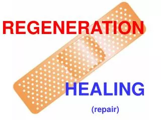
REGENERATION HEALING (repair)
LEARNING OBJECTIVES • Review the normal physiology and concepts of cell proliferation, cell growth, cell “cycle”, and cell differentiation • Understand the basic factors of tissue regeneration • Understand the relationships between cells and their ExtraCellular Matrix (ECM) • Understand the roles of the major players of healing---angiogenesis, growth factors (GFs), and fibrosis • Differentiate 1st & 2nd intention healing
DEFINITIONS: • REGENERATION: Growth of cells to replace lost tissues • HEALING: A reparative tissue response to a wound, inflammation or necrosis, often leads to fibrosis • GRANULATION TISSUE • “ORGANIZING” INFLAMATION
REGENERATION • Replacement of lost structures • Is dependent on the type of normal turnover the original tissue has • Can be differentiated from “compensatory” growth
HEALING (repair) • Needs a wound, inflammatory process, or necrosis • Many disease appearances anatomically are the result of “healing” such as atherosclerosis • Often ends with a scar • Fibrosis, as one of the 3 possible outcomes of inflammation, follows “healing” • Requires a connective tissue “scaffold” • Fibrosis occurs in proportion to the damage of the ECM
Cell Population Fates • PROLIFERATION • Hormonal, especially steroid hormones • eg., EPO, CSF • DIFFERENTIATION* • UNIDIRECTIONAL, GAIN and LOSS • APOPTOSIS *One of the most KEY concepts in neoplasia
ECTODERM MESODERM ENTODERM
CELL CYCLE • G0 • Quiescent (not a very long or dominent phase) • G1 • PRE-synthetic, but cell GROWTH taking place • S • Cells which have continuous “turnover” have longer, or larger S-phases, i.e., DNA synthesis • S-phase of TUMOR CELLS can be prognostic • G2 • PRE-mitotic • M (Mitotic:, P,M,A,T, Cytokinesis)
CELL TYPES • Labile: eg., marrow, GI • Quiescent: liver, kidney • NON-mitotic: neuron, striated muscle
STEM CELLS(TOTIPOTENTIAL*) • EMBRYONIC • ADULT
EMBRYONICSTEM CELLS • DIFFERENTIATION • KNOCKOUT MICE (mice raised with specific gene defects) • REPOPULATION OF DAMAGED TISSUES, in research
ADULTSTEM CELLS • MARROW (HEMOCYTOBLAST) (hematopoetic stem cells) • NON-MARROW (RESERVE)
ADULT TISSUE DIFFERENTIATION and REGENERATION PARALLELS EMBRYONIC DEVELOPMENT
Growth Factors (GFs) • Polypeptides • Cytokines • LOCOMOTION • CONTRACTILITY • DIFFERENTIATION • ANGIOGENESIS
Growth Factors (GFs) • Epidermal • Transforming (alpha, beta) • Hepatocyte • Vascular Endothelial • Platelet Derived • Fibroblast • Keratinocyte • Cytokines (TNF, IL-1, Interferons)
CELL PLAYERS (source AND targets) • Lymphocytes, especially T-cells • Macrophages • Platelets • Endothelial cells • Fibroblasts • Keratinocytes • “Mesenchymal” cells • Smooth muscle cells
E (Epidermal) GF • Made in platelets, macrophages • Present in saliva, milk, urine, plasma • Acts on keratinocytes to migrate, divide • Acts on fibroblasts to produce “granulation” tissue
T (Transforming) GF-alpha • Made in macrophages, T-cells, keratinocytes • Similar to EGF, also effect on hepatocytes
H (Hepatocyte) GF • Made in “mesenchymal” cells • Proliferation of epithelium, endothelium, hepatocytes • Effect on cell “motility”
VE (Vascular Endothelial) GF • Made in mesenchymal cells • Triggered by HYPOXIA • Increases vascular permeability • Mitogenic for endothelial cells • KEY substance in promoting “granulation” tissue
PD (Platelet Derived) GF • Made in platelets, but also MANY other cell types • Chemotactic for MANY cells • Mitogen for fibroblasts • Angiogenesis • Another KEY player in granulation tissue
F (Fibroblast) GF • Made in MANY cells • Chemotactic and mitogenic, for fibroblasts and keratinocytes • Re-epithelialization • Angiogenesis, wound contraction • Hematopoesis • Cardiac/Skeletal (striated) muscle
T (Transforming) GF-beta • Made in MANY CELLS • Chemotactic for PMNs and MANY other types of cells • Inhibits epithelial cells • Fibrogenic • Anti-Inflammatory
K (Keratinocyte) GF • Made in fibroblasts • Stimulates keratinocytes: • Migration • Proliferation • Differentiation
I (Insulin-like) GF-1 • Made in macrophages, fibroblasts • Stimulates: • Sulfated proteoglycans • Collagen • Keratinocyte migration • Fibroblast proliferation • Action similar to GH (Pituitary Growth Hormone)
TNF (Tumor Necrosis Factor) • Made in macrophages, mast cells, T-cells • Activates macrophages (cachexin) • KEY influence on other cytokines • The MAJOR TNF is TNF-alpha
Interleukins • Made in macrophages, mast cells, T-cells, but also MANY other cells • MANY functions: • Chemotaxis • Angiogenesis • REGULATION of other cytokines
INTERFERONS • Made by lymphocytes, fibroblasts • Activates MACROPHAGES • Inhibits FIBROBLASTS • REGULATES other cytokines
SIGNALING • Autocrine (same cell) • Paracrine (next door neighbor) (many GFs) • Endocrine (far away, delivered by blood, steroid hormones)
TRANSCRIPTION FACTORS HEPATIC REGENERATION TNF IL6 HGF
ExtraCellular Matrix (ECM) • Collagen(s) I-XVIII • Elastin • Fibrillin • CAMs (Cell Adhesion Molecules) • Immunoglobulins,cadherins, integrins, selectins • Proteoglycans • Hyaluronic Acid
ECM • Maintain cell differentiation • “Scaffolding” • Establish microenvironment • Storage of GF’s
Collagen One - bONE (main component of bone) Collagen Two - carTWOlage (main component of cartilage) Collagen Three - reTHREEculate (main component of reticular fibers) Collagen Four - FLOOR - forms the basement membrane
GENETIC COLLAGEN DISORDERS • I OSTEOGENESIS IMPERFECTA, E-D • II ACHONDROGENESIS TYPE II • III VASCULAR EHLERS-DANLOS • V CLASSICAL E-D • IX STICKLER SYNDROME • IV ALPORT SYNDROME • VI BETHLEM MYOPATHY • VII DYSTROPHIC EPIDERMOLYSIS BULLOS. • IX EPIPHYSEAL DYSPLASIAS • XVII GEN. EPIDERMOLYSYS BULLOSA • XV, XVIII KNOBLOCH SYNDROME
DEFINITIONS: • REGENERATION: Growth of cells to replace lost tissues • HEALING: A reparative tissue response to a wound, inflammation or necrosis
HEALING • FOLLOWS INFLAMMATION • PROLIFERATION and MIGRATION of connective tissue cells • ANGIOGENESIS (Neovascularization) • Collagen, other ECM protein synthesis • Tissue Remodeling • Wound contraction • Increase in wound strength (scar = fibrosis)
ANGIOGENESIS(NEOVASCULARIZATION) • From endothelial precursor cells • From PRE-existing vessels • Stimulated/Regulated by GF’s, especially VEGF • Also regulated by ECM proteins • aka, “GRANULATION”, “GRANULATION TISSUE”, “ORGANIZATION”, “ORGANIZING INFLAMMATION”
1st INTENTION Edges lined up 2nd INTENTION Edges NOT lined up Ergo…. More granulation More epithelialization MORE FIBROSIS WOUND HEALING
FIBROSIS/SCARRING • DEPOSITION OF COLLAGEN by FIBROBLASTS • With time (weeks, months, years?) the collagen becomes more dense, ergo, the tissue becomes “STRONGER”
Wound RETARDING factors(LOCAL) • DECREASED Blood supply • Denervation • Local Infection • FB • Hematoma • Mechanical stress • Necrotic tissue